Liu Yang, Yang Faji, Li Jun, Wang Jinglin, Wang Xun, Zhang Yuheng, Yuan Xianwen, Zhu Wei, Shi Xiaolei
Department of Hepatobiliary Surgery, The Affiliated Drum Tower Hospital of Nanjing University Medical School, Nanjing, Jiangsu 210008, China.
Department of Anesthesiology, The Affiliated Drum Tower Hospital of Nanjing University Medical School, Nanjing, Jiangsu 210008, China.
Stem Cells Int. 2018 May 13;2018:7652359. doi: 10.1155/2018/7652359. eCollection 2018.
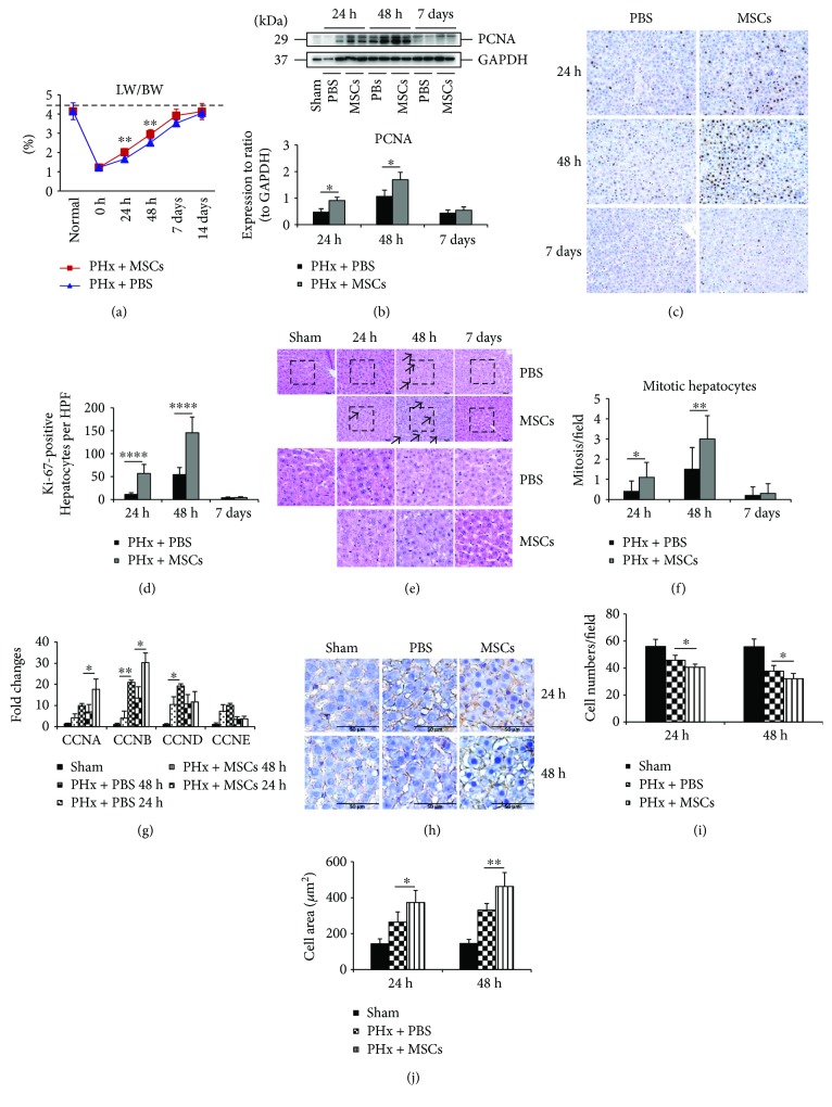
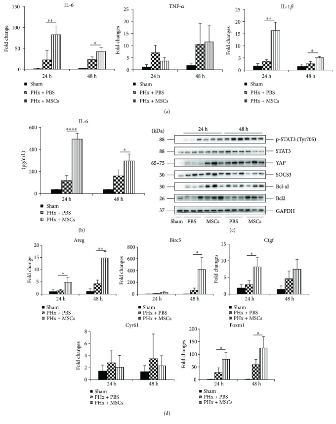
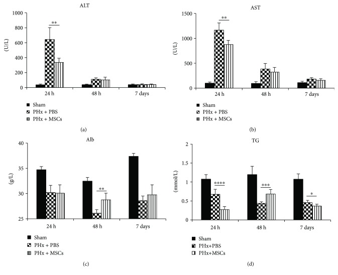
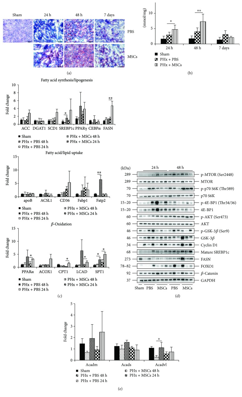
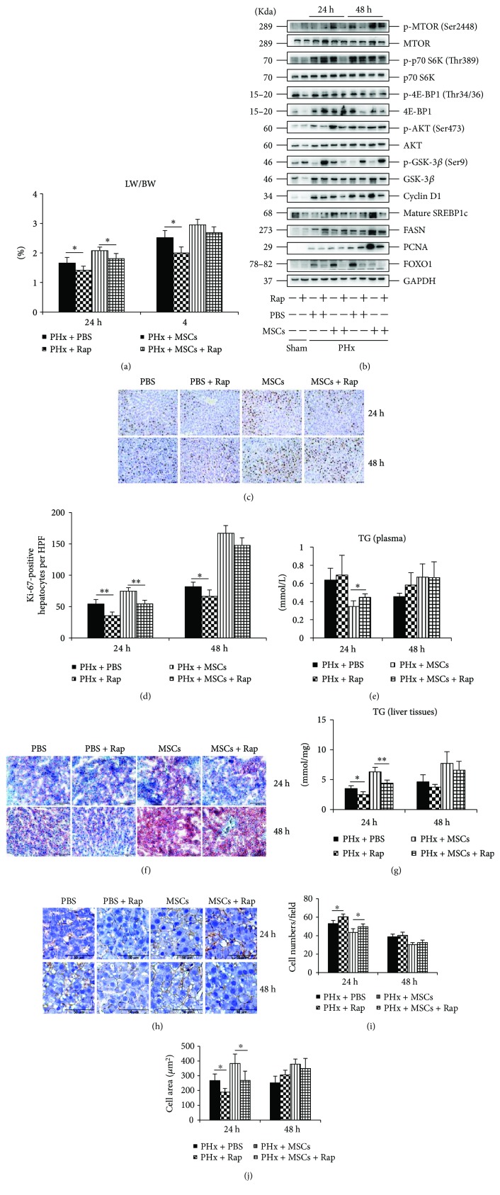
The liver has the potential to regenerate after injury. It is a challenge to improve liver regeneration (LR) after liver resection in clinical practice. Bone morrow-derived mesenchymal stem cells (MSCs) have shown to have a role in various liver diseases. To explore the effects of MSCs on LR, we established a model of 70% partial hepatectomy (PHx). Results revealed that infusion of MSCs could improve LR through enhancing cell proliferation and cell growth during the first 2 days after PHx, and MSCs could also restore liver synthesis function. Infusion of MSCs also improved liver lipid accumulation partly via mechanistic target of rapamycin (mTOR) signaling and enhanced lipid -oxidation support energy for LR. Rapamycin-induced inhibition of mTOR decreased liver lipid accumulation at 24 h after PHx, leading to impaired LR. And after infusion of MSCs, a proinflammatory environment formed in the liver, evidenced by increased expression of IL-6 and IL-1, and thus the STAT3 and Hippo-YAP pathways were activated to improve cell proliferation. Our results demonstrated the function of MSCs on LR after PHx and provided new evidence for stem cell therapy of liver diseases.
肝脏在损伤后具有再生潜力。在临床实践中,提高肝切除术后的肝再生(LR)是一项挑战。骨髓来源的间充质干细胞(MSCs)已被证明在各种肝脏疾病中发挥作用。为了探究MSCs对LR的影响,我们建立了70%部分肝切除术(PHx)模型。结果显示,输注MSCs可通过在PHx后的头2天增强细胞增殖和细胞生长来改善LR,并且MSCs还可恢复肝脏合成功能。输注MSCs还部分通过雷帕霉素靶蛋白(mTOR)信号通路改善肝脏脂质蓄积,并增强脂质氧化为LR提供能量支持。雷帕霉素诱导的mTOR抑制在PHx后24小时降低了肝脏脂质蓄积,导致LR受损。并且在输注MSCs后,肝脏中形成了促炎环境,表现为IL-6和IL-1表达增加,因此STAT3和Hippo-YAP信号通路被激活以改善细胞增殖。我们的结果证明了MSCs在PHx后对LR的作用,并为肝脏疾病的干细胞治疗提供了新证据。