Panuccio Gabriella, Colombi Ilaria, Chiappalone Michela
Department of Neuroscience and Brain Technologies, Istituto Italiano di Tecnologia;
Department of Neuroscience and Brain Technologies, Istituto Italiano di Tecnologia.
J Vis Exp. 2018 May 15(135):57548. doi: 10.3791/57548.
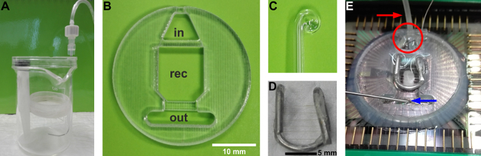
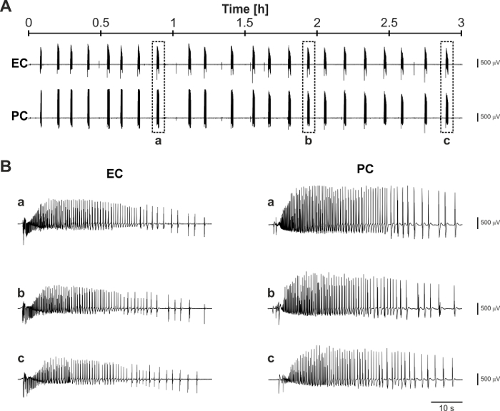
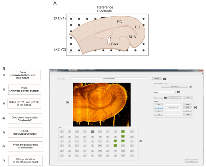
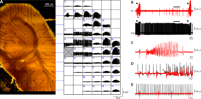
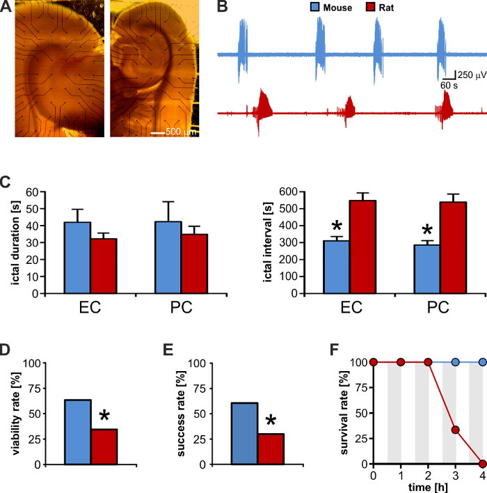
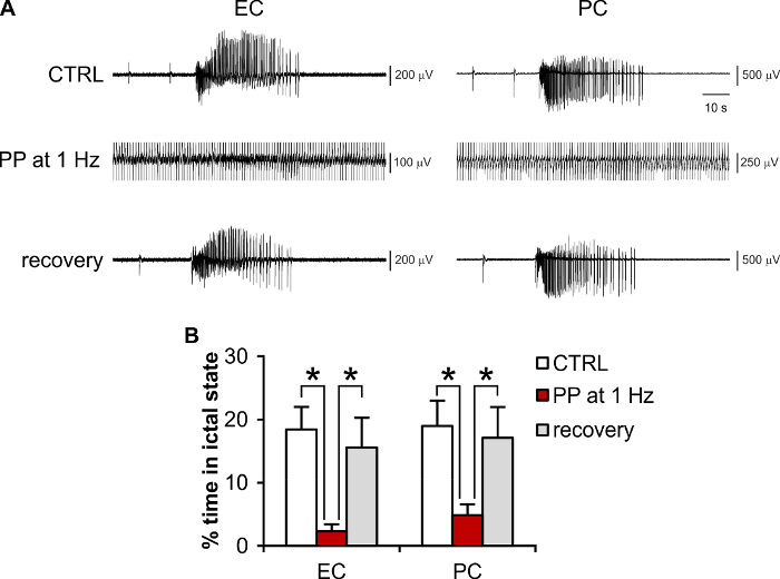
Temporal lobe epilepsy (TLE) is the most common partial complex epileptic syndrome and the least responsive to medications. Deep brain stimulation (DBS) is a promising approach when pharmacological treatment fails or neurosurgery is not recommended. Acute brain slices coupled to microelectrode arrays (MEAs) represent a valuable tool to study neuronal network interactions and their modulation by electrical stimulation. As compared to conventional extracellular recording techniques, they provide the added advantages of a greater number of observation points and a known inter-electrode distance, which allow studying the propagation path and speed of electrophysiological signals. However, tissue oxygenation may be greatly impaired during MEA recording, requiring a high perfusion rate, which comes at the cost of decreased signal-to-noise ratio and higher oscillations in the experimental temperature. Electrical stimulation further stresses the brain tissue, making it difficult to pursue prolonged recording/stimulation epochs. Moreover, electrical modulation of brain slice activity needs to target specific structures/pathways within the brain slice, requiring that electrode mapping be easily and quickly performed live during the experiment. Here, we illustrate how to perform the recording and electrical modulation of 4-aminopyridine (4AP)-induced epileptiform activity in rodent brain slices using planar MEAs. We show that the brain tissue obtained from mice outperforms rat brain tissue and is thus better suited for MEA experiments. This protocol guarantees the generation and maintenance of a stable epileptiform pattern that faithfully reproduces the electrophysiological features observed with conventional field potential recording, persists for several hours, and outlasts sustained electrical stimulation for prolonged epochs. Tissue viability throughout the experiment is achieved thanks to the use of a small-volume custom recording chamber allowing for laminar flow and quick solution exchange even at low (1 mL/min) perfusion rates. Quick MEA mapping for real-time monitoring and selection of stimulating electrodes is performed by a custom graphic user interface (GUI).
颞叶癫痫(TLE)是最常见的部分性复杂性癫痫综合征,对药物治疗反应最差。当药物治疗失败或不建议进行神经外科手术时,深部脑刺激(DBS)是一种很有前景的方法。与微电极阵列(MEA)耦合的急性脑片是研究神经元网络相互作用及其电刺激调制的宝贵工具。与传统的细胞外记录技术相比,它们具有更多观察点和已知电极间距的额外优势,这有助于研究电生理信号的传播路径和速度。然而,在MEA记录过程中,组织氧合可能会受到极大损害,需要高灌注速率,这会导致信噪比降低和实验温度出现更高的波动。电刺激会进一步给脑组织带来压力,使得难以进行长时间的记录/刺激时段。此外,脑片活动的电调制需要针对脑片内的特定结构/通路,这要求在实验过程中能够轻松快速地实时进行电极映射。在这里,我们说明了如何使用平面MEA在啮齿动物脑片中进行4-氨基吡啶(4AP)诱导的癫痫样活动的记录和电调制。我们表明,从小鼠获得的脑组织优于大鼠脑组织,因此更适合进行MEA实验。该方案保证了稳定癫痫样模式的产生和维持,该模式忠实地再现了传统场电位记录中观察到的电生理特征,持续数小时,并在长时间的持续电刺激后仍能持续存在。由于使用了小体积定制记录室,即使在低灌注速率(1 mL/分钟)下也能实现层流和快速溶液交换,从而在整个实验过程中实现了组织活力。通过定制图形用户界面(GUI)进行快速MEA映射,以实时监测和选择刺激电极。