Neurological Clinical Research Institute, Massachusetts General Hospital, Boston, Massachusetts.
Department of Psychiatry, Jigme Dorji Wangchuck National Referral Hospital, Thimphu, Bhutan.
Am J Trop Med Hyg. 2018 Aug;99(2):482-488. doi: 10.4269/ajtmh.17-0943. Epub 2018 Jun 7.
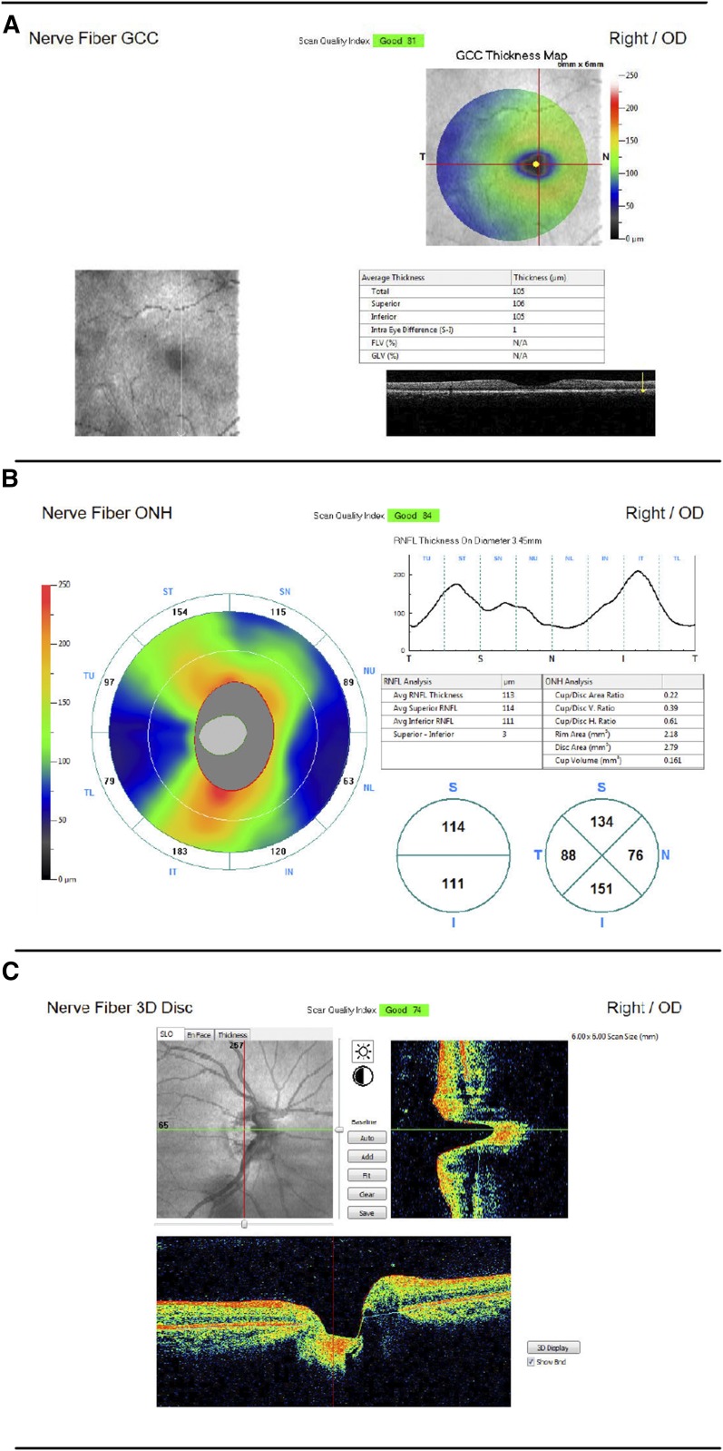
The retina shares embryological derivation with the brain and may provide a new measurement of overall growth status, especially useful in resource-limited settings. Optical coherence tomography (OCT) provides detailed quantification of retinal structures. We enrolled community-dwelling children ages 3-11 years old in Siaya, Kenya and Thimphu, Bhutan in 2016. We measured head circumference (age < 5 years only), height, and weight, and standardized these by age and gender. Research staff performed OCT (; Optovue, Inc., Fremont, CA), measuring the peripapillary retinal nerve fiber layer (RNFL) and macular ganglion cell complex (GCC) thicknesses. A neuro-ophthalmologist performed quality control for centration, motion artifact, and algorithm-derived quality scores. Generalized estimating equations were used to determine the relationship between anthropometric and retinal measurements. Two hundred and fifty-eight children (139 females, average age 6.4 years) successfully completed at least one retinal scan, totaling 1,048 scans. Nine hundred and twenty-two scans (88.0%) were deemed usable. Fifty-three of the 258 children (20.5%) were able to complete all six scans. Kenyan children had a thinner average GCC ( < 0.001) than Bhutanese children after adjustment for age and gender, but not RNFL ( = 0.70). In models adjusting for age, gender, and study location, none of standardized height, weight, and body mass index (BMI) were statistically significantly associated with RNFL or GCC. We determined that OCT is feasible in some children in resource-limited settings, particularly those > 4 years old, using the device. We found no evidence for GCC or RNFL as a proxy for height-, weight-, or BMI-for-age. The variation in mean GCC thickness in Asian versus African children warrants further investigation.
视网膜与大脑具有胚胎学上的同源性,可能提供整体生长状况的新测量指标,尤其在资源有限的环境中非常有用。光学相干断层扫描(OCT)可对视网膜结构进行详细的定量分析。我们于 2016 年在肯尼亚的锡亚亚和不丹的廷布招募了年龄在 3-11 岁的社区居住儿童。我们测量了头围(仅在年龄 < 5 岁的儿童中进行)、身高和体重,并按年龄和性别进行了标准化。研究人员进行了 OCT(Optovue,Inc.,加利福尼亚州弗里蒙特)检查,测量了视盘周围视网膜神经纤维层(RNFL)和黄斑神经节细胞复合体(GCC)的厚度。神经眼科医生对中心定位、运动伪影和算法衍生的质量分数进行了质量控制。使用广义估计方程来确定人体测量学和视网膜测量之间的关系。共有 258 名儿童(139 名女性,平均年龄 6.4 岁)成功完成了至少一次视网膜扫描,共获得 1048 次扫描结果。922 次扫描(88.0%)被认为是可用的。在 258 名儿童中,有 53 名(20.5%)能够完成所有 6 次扫描。在调整年龄和性别后,肯尼亚儿童的平均 GCC 较薄(<0.001),但 RNFL 没有差异(=0.70)。在调整年龄、性别和研究地点的模型中,标准化身高、体重和体重指数(BMI)均与 RNFL 或 GCC 无统计学关联。我们确定,在资源有限的环境中,使用该设备对一些儿童进行 OCT 检查是可行的,特别是 4 岁以上的儿童。我们没有发现 GCC 或 RNFL 可以作为身高、体重或 BMI 与年龄相关的替代指标。亚洲儿童与非洲儿童之间平均 GCC 厚度的差异值得进一步研究。