IEEE J Biomed Health Inform. 2018 Jul;22(4):1168-1176. doi: 10.1109/JBHI.2017.2762520.
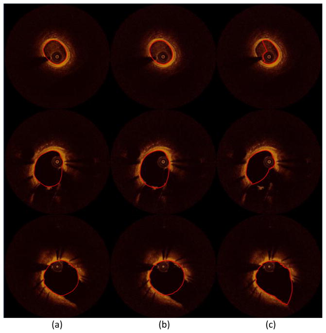
We present a novel and time-efficient method for intracoronary lumen detection, which produces three-dimensional (3-D) coronary arteries using optical coherence tomographic (OCT) images. OCT images are acquired for multiple patients and longitudinal cross-section (LOCS) images are reconstructed using different acquisition angles. The lumen contours for each LOCS image are extracted and translated to 2-D cross-sectional images. Using two angiographic projections, the centerline of the coronary vessel is reconstructed in 3-D, and the detected 2-D contours are transformed to 3-D and placed perpendicular to the centerline. To validate the proposed method, 613 manual annotations from medical experts were used as gold standard. The 2-D detected contours were compared with the annotated contours, and the 3-D reconstructed models produced using the detected contours were compared to the models produced by the annotated contours. Wall shear stress (WSS), as dominant hemodynamics factor, was calculated using computational fluid dynamics and 844 consecutive 2-mm segments of the 3-D models were extracted and compared with each other. High Pearson's correlation coefficients were obtained for the lumen area (r = 0.98) and local WSS (r = 0.97) measurements, while no significant bias with good limits of agreement was shown in the Bland-Altman analysis. The overlapping and nonoverlapping areas ratio between experts' annotations and presented method was 0.92 and 0.14, respectively. The proposed computer-aided lumen extraction and 3-D vessel reconstruction method is fast, accurate, and likely to assist in a number of research and clinical applications.
我们提出了一种新颖的、高效的方法来检测冠状动脉内腔,该方法使用光学相干断层扫描(OCT)图像生成三维(3-D)冠状动脉。对多个患者进行 OCT 图像采集,并使用不同的采集角度重建纵向横截面(LOCS)图像。提取每个 LOCS 图像的内腔轮廓,并将其转换为 2-D 横截面图像。使用两个血管造影投影,在 3-D 中重建冠状动脉的中心线,并将检测到的 2-D 轮廓转换为 3-D 并垂直于中心线放置。为了验证所提出的方法,使用 613 条来自医学专家的手动注释作为金标准。将 2-D 检测到的轮廓与注释轮廓进行比较,并将使用检测到的轮廓生成的 3-D 重建模型与使用注释轮廓生成的模型进行比较。壁面剪切应力(WSS)作为主导的血液动力学因素,使用计算流体动力学进行计算,并从 3-D 模型中提取 844 个连续的 2-mm 片段进行相互比较。内腔面积(r = 0.98)和局部 WSS(r = 0.97)测量得到了较高的 Pearson 相关系数,而 Bland-Altman 分析显示没有显著的偏差和良好的一致性范围。专家注释和提出的方法之间的重叠和非重叠区域比例分别为 0.92 和 0.14。该方法快速、准确,有望辅助许多研究和临床应用。