Sargolzaei Aval Fereydoon, Arab Mohammad Reza, Sargolzaei Narjes, Noushadi Fateme, Eteghadi Abdolsamad, Keykhaei Asadollah, Sargolzaei Aval Foroug, Hedayat Pour Azim
Associate Professor, Cellular and Molecular Research Center, Department of Anatomical Sciences, School of Medicine, Zahedan University of Medical Sciences, Zahedan, Iran.
Professor, Cellular and Molecular Research Center, Department of Anatomical Sciences, School of Medicine, Zahedan University of Medical Sciences, Zahedan, Iran.
J Dent (Tehran). 2018 Mar;15(2):86-96.
The healing of bone defects in the craniofacial region is an important clinical issue. We aimed to compare the effects of octacalcium phosphate (OCP) and the combination of OCP/gelatin (OCP/Gel) on calvarial bone regeneration in rats.
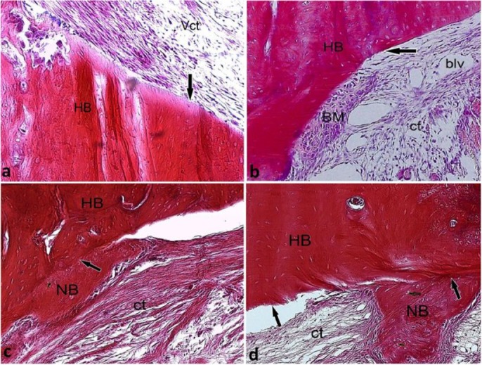
In this study, 72 male Sprague Dawley rats were randomly assigned to the OCP (n=24), OCP/Gel (n=24), and control groups (n=24). Lesions with a diameter of 9 mm were created in the parietal bone and were filled with 9-mg OCP and OCP/Gel disks. In the control group, no substance was implanted in the defect. Sampling was performed on days 10, 14, 21, and 28 after the implantation. After tissue processing, 5-μm sections were prepared and stained by hematoxylin and eosin (H&E) stain. The sections were studied, and the volume fraction of the newly formed bone was assessed by Kruskal-Wallis test at a significance level of 0.05.
In the experimental groups, new bone formation was detected at the margins of the defects 10 days after the implantation. With the progression of the healing process, the newly formed bone covered greater areas of the defects and developed a more mature structure. In the control group, the defects were primarily filled with a dense connective tissue with small islands of new bone. The results of histomorphometric assessments showed that the volume of the newly formed bone in the experimental groups had a significant statistical difference with that in the control group (P<0.001).
The OCP/Gel composite can be useful in the healing process of calvarial bone defects.
颅面部骨缺损的愈合是一个重要的临床问题。我们旨在比较磷酸八钙(OCP)和OCP/明胶组合(OCP/Gel)对大鼠颅骨再生的影响。
在本研究中,72只雄性Sprague Dawley大鼠被随机分为OCP组(n = 24)、OCP/Gel组(n = 24)和对照组(n = 24)。在顶骨上制造直径为9 mm的缺损,并填充9 mg的OCP和OCP/Gel盘。对照组在缺损处不植入任何物质。在植入后第10、14、21和28天进行取样。组织处理后,制备5μm切片并用苏木精和伊红(H&E)染色。对切片进行研究,并通过Kruskal-Wallis检验在显著性水平为0.05时评估新形成骨的体积分数。
在实验组中,植入后10天在缺损边缘检测到新骨形成。随着愈合过程的进展,新形成的骨覆盖了缺损的更大区域并发育出更成熟的结构。在对照组中,缺损主要填充有致密结缔组织和小的新骨岛。组织形态计量学评估结果表明,实验组中新形成骨的体积与对照组有显著统计学差异(P < 0.001)。
OCP/Gel复合材料可用于颅骨缺损的愈合过程。