Munich Center for Integrated Protein Science (CIPSM) at the Department of Biology I, Microbiology, Ludwig-Maximilians-Universität München, Martinsried, Germany.
Max Plank Institute for Biochemistry, Martinsried, Germany.
Sci Rep. 2018 Jul 4;8(1):10137. doi: 10.1038/s41598-018-28472-0.
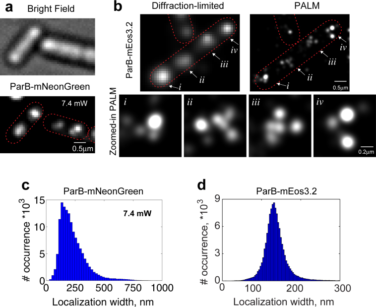
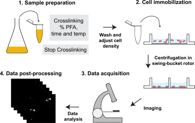
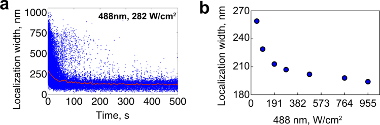
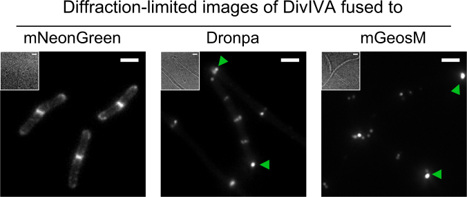
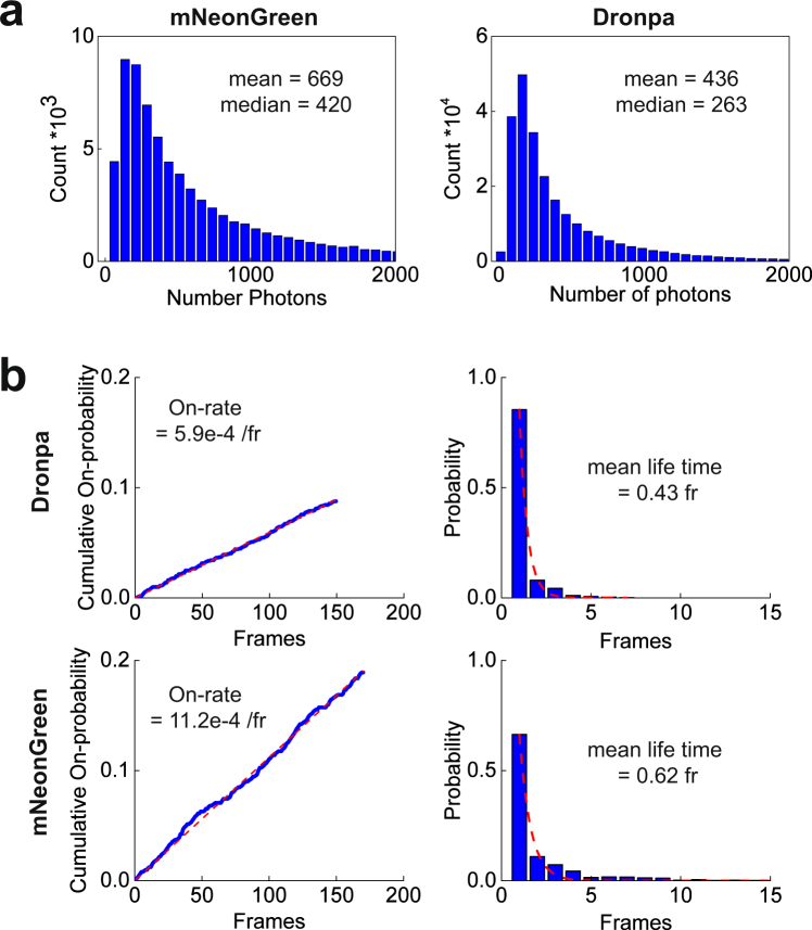
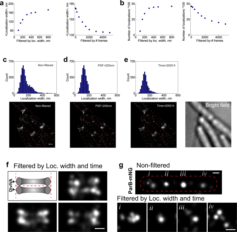
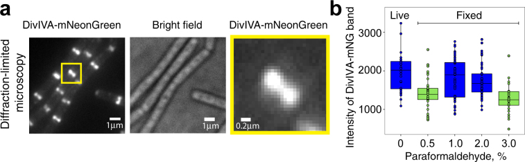
mNeonGreen fluorescent protein is capable of photo-switching, hence in principle applicable for super-resolution imaging. However, difficult-to-control blinking kinetics that lead to simultaneous emission of multiple nearby mNeonGreen molecules impedes its use for PALM. Here, we determined the on- and off- switching rate and the influence of illumination power on the simultaneous emission. Increasing illumination power reduces the probability of simultaneous emission, but not enough to generate high quality PALM images. Therefore, we introduce a simple data post-processing step that uses temporal and spatial information of molecule localizations to further reduce artifacts arising from simultaneous emission of nearby emitters. We also systematically evaluated various sample preparation steps to establish an optimized protocol to preserve cellular morphology and fluorescence signal. In summary, we propose a workflow for super-resolution imaging with mNeonGreen based on optimization of sample preparation, data acquisition and simple post-acquisition data processing. Application of our protocol enabled us to resolve the expected double band of bacterial cell division protein DivIVA, and to visualize that the chromosome organization protein ParB organized into sub-clusters instead of the typically observed diffraction-limited foci. We expect that our workflow allows a broad use of mNeonGreen for super-resolution microscopy, which is so far difficult to achieve.
mNeonGreen 荧光蛋白能够光开关,因此原则上适用于超分辨率成像。然而,难以控制的闪烁动力学导致多个附近的 mNeonGreen 分子同时发射,这阻碍了其在 PALM 中的应用。在这里,我们确定了开/关转换率以及光照强度对同时发射的影响。增加光照强度会降低同时发射的概率,但不足以产生高质量的 PALM 图像。因此,我们引入了一个简单的数据后处理步骤,该步骤使用分子定位的时间和空间信息来进一步减少来自附近发射器同时发射的伪影。我们还系统地评估了各种样品制备步骤,以建立一种优化的方案来保留细胞形态和荧光信号。总之,我们提出了一种基于优化样品制备、数据采集和简单后处理的使用 mNeonGreen 进行超分辨率成像的工作流程。我们的方案的应用使我们能够分辨出细菌细胞分裂蛋白 DivIVA 的预期双带,并观察到染色体组织蛋白 ParB 组织成亚簇,而不是通常观察到的衍射受限焦点。我们期望我们的工作流程能够广泛应用于超分辨率显微镜,这在目前是很难实现的。