• 文献检索
  • 文档翻译
  • 深度研究
  • 学术资讯
  • Suppr Zotero 插件Zotero 插件
  • 邀请有礼
  • 套餐&价格
  • 历史记录
应用&插件
Suppr Zotero 插件Zotero 插件浏览器插件Mac 客户端Windows 客户端微信小程序
定价
高级版会员购买积分包购买API积分包
服务
文献检索文档翻译深度研究API 文档MCP 服务
关于我们
关于 Suppr公司介绍联系我们用户协议隐私条款
关注我们

Suppr 超能文献

核心技术专利:CN118964589B侵权必究
粤ICP备2023148730 号-1Suppr @ 2026

文献检索

告别复杂PubMed语法,用中文像聊天一样搜索,搜遍4000万医学文献。AI智能推荐,让科研检索更轻松。

立即免费搜索

文件翻译

保留排版,准确专业,支持PDF/Word/PPT等文件格式,支持 12+语言互译。

免费翻译文档

深度研究

AI帮你快速写综述,25分钟生成高质量综述,智能提取关键信息,辅助科研写作。

立即免费体验

利用电子断层扫描和能量色散X射线光谱对高温氧化进行定量表征。

Quantitative characterization of high temperature oxidation using electron tomography and energy-dispersive X-ray spectroscopy.

作者信息

Zhou Jihan, Taylor Matthew, Melinte Georgian A, Shahani Ashwin J, Dharmawardhana Chamila C, Heinz Hendrik, Voorhees Peter W, Perepezko John H, Bustillo Karen, Ercius Peter, Miao Jianwei

机构信息

Department of Physics and Astronomy and California NanoSystems Institute, University of California, Los Angeles, CA, 90095, USA.

Department of Materials Science and Engineering, University of Wisconsin-Madison, Madison, WI, 53706, USA.

出版信息

Sci Rep. 2018 Jul 6;8(1):10239. doi: 10.1038/s41598-018-28348-3.

DOI:10.1038/s41598-018-28348-3
PMID:29980704
原文链接:https://pmc.ncbi.nlm.nih.gov/articles/PMC6035195/
Abstract

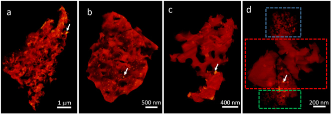
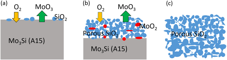
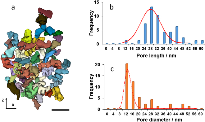
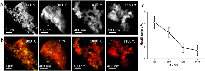
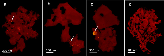
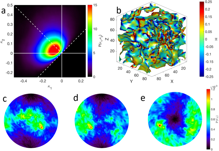
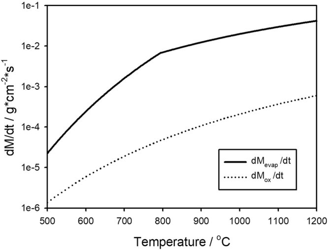
We report quantitative characterization of the high temperature oxidation process by using electron tomography and energy-dispersive X-ray spectroscopy. As a proof of principle, we performed 3D imaging of the oxidation layer of a model system (MoSi) at nanoscale resolution with elemental specificity and probed the oxidation kinetics as a function of the oxidation time and the elevated temperature. Our tomographic reconstructions provide detailed 3D structural information of the surface oxidation layer of the MoSi system, revealing the evolution of oxidation behavior of MoSi from early stage to mature stage. Based on the relative rate of oxidation of MoSi, the volatilization rate of MoO and reactive molecular dynamics simulations, we propose a model to explain the mechanism of the formation of the porous silica structure during the oxidation process of MoSi. We expect that this 3D quantitative characterization method can be applied to other material systems to probe their structure-property relationships in different environments.

摘要

我们通过电子断层扫描和能量色散X射线光谱法报告了高温氧化过程的定量表征。作为原理验证,我们以纳米级分辨率对模型系统(MoSi)的氧化层进行了具有元素特异性的三维成像,并探究了氧化动力学与氧化时间和升高温度的函数关系。我们的断层扫描重建提供了MoSi系统表面氧化层的详细三维结构信息,揭示了MoSi从早期到成熟阶段氧化行为的演变。基于MoSi的相对氧化速率、MoO的挥发速率和反应性分子动力学模拟,我们提出了一个模型来解释MoSi氧化过程中多孔二氧化硅结构的形成机制。我们期望这种三维定量表征方法能够应用于其他材料系统,以探究它们在不同环境中的结构-性能关系。

https://cdn.ncbi.nlm.nih.gov/pmc/blobs/fff2/6035195/5a7e7af09ac6/41598_2018_28348_Fig7_HTML.jpg
https://cdn.ncbi.nlm.nih.gov/pmc/blobs/fff2/6035195/5e83e9253ad2/41598_2018_28348_Fig1_HTML.jpg
https://cdn.ncbi.nlm.nih.gov/pmc/blobs/fff2/6035195/157d2232d65d/41598_2018_28348_Fig2_HTML.jpg
https://cdn.ncbi.nlm.nih.gov/pmc/blobs/fff2/6035195/1b9b498afb97/41598_2018_28348_Fig3_HTML.jpg
https://cdn.ncbi.nlm.nih.gov/pmc/blobs/fff2/6035195/defc89cc9cd3/41598_2018_28348_Fig4_HTML.jpg
https://cdn.ncbi.nlm.nih.gov/pmc/blobs/fff2/6035195/483ab49e06b5/41598_2018_28348_Fig5_HTML.jpg
https://cdn.ncbi.nlm.nih.gov/pmc/blobs/fff2/6035195/2c1dbb91b788/41598_2018_28348_Fig6_HTML.jpg
https://cdn.ncbi.nlm.nih.gov/pmc/blobs/fff2/6035195/5a7e7af09ac6/41598_2018_28348_Fig7_HTML.jpg

相似文献

1
Quantitative characterization of high temperature oxidation using electron tomography and energy-dispersive X-ray spectroscopy.利用电子断层扫描和能量色散X射线光谱对高温氧化进行定量表征。
Sci Rep. 2018 Jul 6;8(1):10239. doi: 10.1038/s41598-018-28348-3.
2
Formation of MoSi/MoSi-MgAlO Composites via Self-Propagating High-Temperature Synthesis.自蔓延高温合成法制备 MoSi/MoSi-MgAlO 复合材料。
Molecules. 2019 Dec 24;25(1):83. doi: 10.3390/molecules25010083.
3
Energy Dispersive X-ray Tomography for 3D Elemental Mapping of Individual Nanoparticles.用于单个纳米颗粒三维元素映射的能量色散X射线断层扫描技术。
J Vis Exp. 2016 Jul 5(113):52815. doi: 10.3791/52815.
4
Direct observation of incommensurate structure in MoSi.对MoSi中非公度结构的直接观测。
Acta Crystallogr A Found Adv. 2016 Nov 1;72(Pt 6):660-666. doi: 10.1107/S2053273316012286. Epub 2016 Oct 3.
5
3D nanoscale elemental mapping of precipitates in steel: Evaluation of analytical electron tomography and comparison to atom probe tomography.钢中析出物的三维纳米级元素映射:分析电子断层扫描的评估及与原子探针断层扫描的比较。
Micron. 2022 May;156:103233. doi: 10.1016/j.micron.2022.103233. Epub 2022 Mar 5.
6
Multiscale Characterization of an Oxide Scale Formed on the Creep-Resistant ATI 718Plus Superalloy during High-Temperature Oxidation.抗蠕变ATI 718Plus高温合金在高温氧化过程中形成的氧化皮的多尺度表征
Materials (Basel). 2021 Oct 23;14(21):6327. doi: 10.3390/ma14216327.
7
In Situ Industrial Bimetallic Catalyst Characterization using Scanning Transmission Electron Microscopy and X-ray Absorption Spectroscopy at One Atmosphere and Elevated Temperature.在一个大气压和高温下使用扫描透射电子显微镜和X射线吸收光谱对原位工业双金属催化剂进行表征
Chemphyschem. 2017 Aug 18;18(16):2151-2156. doi: 10.1002/cphc.201700425. Epub 2017 Jul 4.
8
Extreme Ultraviolet Reflection-Absorption Spectroscopy: Probing Dynamics at Surfaces from a Molecular Perspective.极端紫外反射-吸收光谱学:从分子角度探究表面动力学。
Acc Chem Res. 2022 Mar 15;55(6):893-903. doi: 10.1021/acs.accounts.1c00765. Epub 2022 Mar 3.
9
Temperature-Dependent Oxidation Behavior of Silicon Carbide Surface: Reactive Molecular Dynamics Simulations.碳化硅表面的温度依赖性氧化行为:反应分子动力学模拟
ACS Appl Mater Interfaces. 2024 Oct 3. doi: 10.1021/acsami.4c12392.
10
Multi-scale 3D characterization of long period stacking ordered structure in Mg-Zn-Gd cast alloys.Mg-Zn-Gd铸造合金中长周期堆垛有序结构的多尺度三维表征
Microscopy (Oxf). 2014 Nov;63 Suppl 1:i25-i26. doi: 10.1093/jmicro/dfu068.

本文引用的文献

1
GENFIRE: A generalized Fourier iterative reconstruction algorithm for high-resolution 3D imaging.GENFIRE:一种用于高分辨率 3D 成像的广义傅里叶迭代重建算法。
Sci Rep. 2017 Sep 5;7(1):10409. doi: 10.1038/s41598-017-09847-1.
2
Deciphering chemical order/disorder and material properties at the single-atom level.解析单原子层次的化学有序/无序和材料性质。
Nature. 2017 Feb 1;542(7639):75-79. doi: 10.1038/nature21042.
3
Atomic electron tomography: 3D structures without crystals.原子电子断层扫描技术:无需晶体的三维结构。
Science. 2016 Sep 23;353(6306). doi: 10.1126/science.aaf2157.
4
Nanomaterial datasets to advance tomography in scanning transmission electron microscopy.推进扫描透射电子显微镜断层成像的纳米材料数据集。
Sci Data. 2016 Jun 7;3:160041. doi: 10.1038/sdata.2016.41.
5
Simulations of inorganic-bioorganic interfaces to discover new materials: insights, comparisons to experiment, challenges, and opportunities.模拟无机-生物有机界面以发现新材料:见解、与实验的比较、挑战和机遇。
Chem Soc Rev. 2016 Jan 21;45(2):412-48. doi: 10.1039/c5cs00890e. Epub 2016 Jan 11.
6
Formation of bimetallic clusters in superfluid helium nanodroplets analysed by atomic resolution electron tomography.通过原子分辨率电子断层扫描分析超流氦纳米液滴中双金属团簇的形成。
Nat Commun. 2015 Oct 28;6:8779. doi: 10.1038/ncomms9779.
7
Three-dimensional coordinates of individual atoms in materials revealed by electron tomography.电子断层扫描揭示材料中单个原子的三维坐标。
Nat Mater. 2015 Nov;14(11):1099-103. doi: 10.1038/nmat4426. Epub 2015 Sep 21.
8
A model based iterative reconstruction algorithm for high angle annular dark field-scanning transmission electron microscope (HAADF-STEM) tomography.一种基于模型的迭代重建算法,用于高角环形暗场扫描透射电子显微镜(HAADF-STEM)断层扫描。
IEEE Trans Image Process. 2013 Nov;22(11):4532-44. doi: 10.1109/TIP.2013.2277784. Epub 2013 Aug 8.
9
Three-dimensional elemental mapping at the atomic scale in bimetallic nanocrystals.在双金属纳米晶体中进行原子级别的三维元素映射。
Nano Lett. 2013 Sep 11;13(9):4236-41. doi: 10.1021/nl401945b. Epub 2013 Aug 20.
10
Three-dimensional imaging of dislocations in a nanoparticle at atomic resolution.在原子分辨率下对纳米粒子中位错的三维成像。
Nature. 2013 Apr 4;496(7443):74-7. doi: 10.1038/nature12009. Epub 2013 Mar 27.