Department of Transdisciplinary Studies, Seoul National University, Seoul, Republic of Korea.
Department of Nuclear Medicine, Seoul National University Bundang Hospital, Seongnam, Republic of Korea.
Sci Rep. 2018 Jul 24;8(1):11122. doi: 10.1038/s41598-018-29424-4.
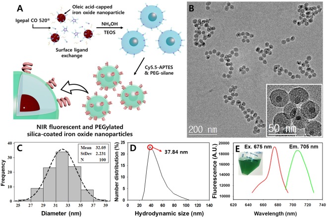
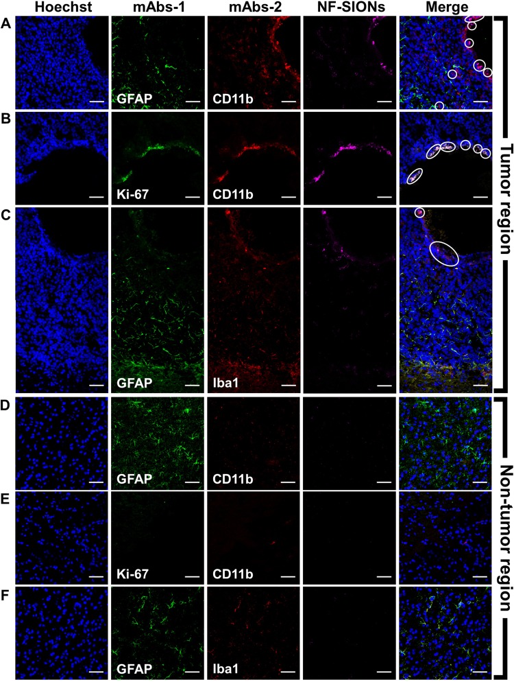
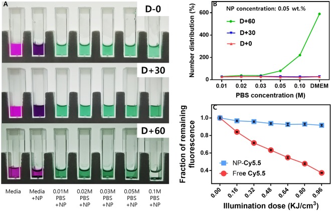
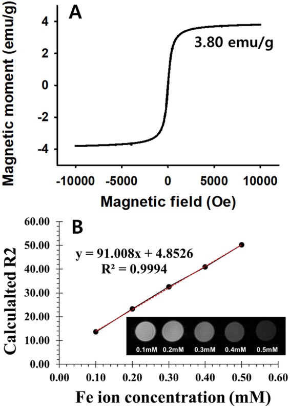
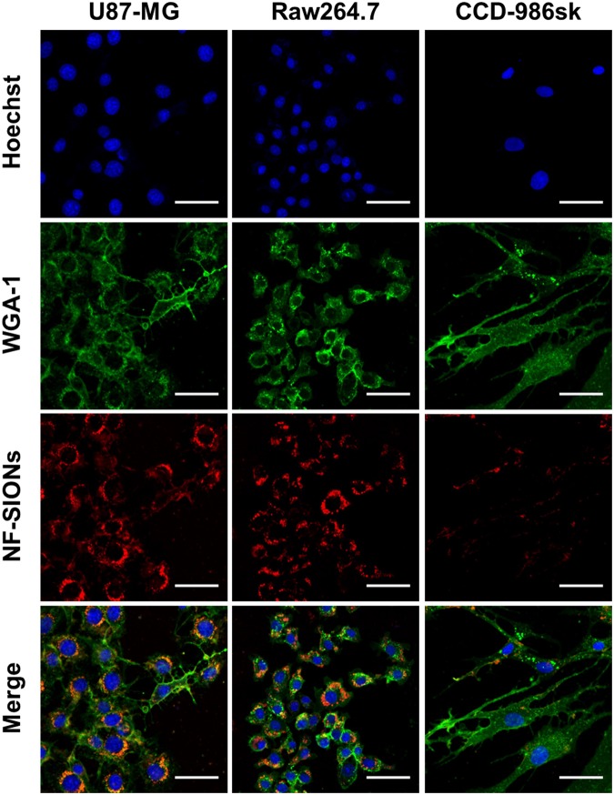
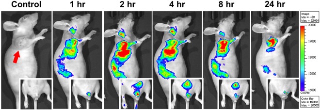
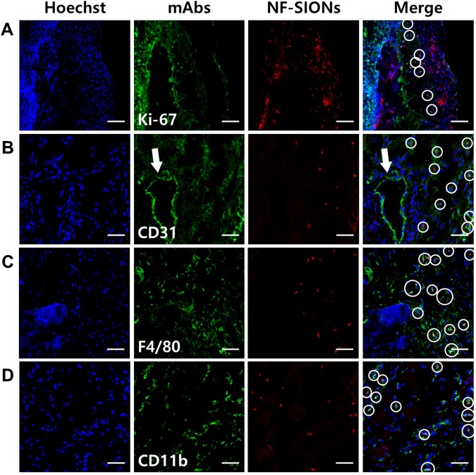
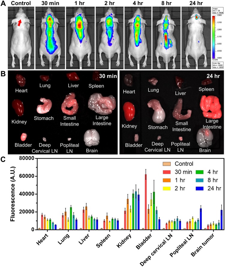
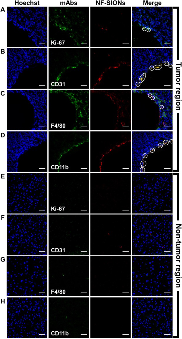
Glioblastoma multiforme (GBM) is the most aggressive and lethal type of human brain cancer. Surgery is a current gold standard for GBM treatment but the complete surgical resection of GBM is almost impossible due to their diffusive characteristics into surrounded normal brain tissues. There is an urgent need to develop a sensitive imaging tool for accurate delineation of GBM in the operating room to guide surgeons. Here we illustrate the feasibility of using near-infrared fluorescent silica coated iron oxide nanoparticles (NF-SIONs) with high water dispersion capacity and strong fluorescence stability for intraoperative imaging of GBM by targeting tumor-associated macrophages. Abundant macrophage infiltration is a key feature of GBM margins and it is well associated with poor prognosis. We synthesized NF-SIONs of about 37 nm to maximize endocytosis activity for macrophage uptake. The NF-SIONs selectively visualized tumor-associated macrophage populations by in vitro live-cell imaging and in vivo fluorescence imaging. In the orthotopic GBM xenograft models, the NF-SIONs could successfully penetrate blood-brain barrier and delineated tumor burden specifically. Taken together, this study showcased the potential applications in GBM treatment for improved intraoperative staging and more radical surgery as well as dual modality benefit in order to circumvent previous clinical failure.
多形性胶质母细胞瘤(GBM)是最具侵袭性和致命性的人类脑癌。手术是目前治疗 GBM 的金标准,但由于其扩散到周围正常脑组织的特性,GBM 的完全手术切除几乎是不可能的。迫切需要开发一种敏感的成像工具,以便在手术室中准确描绘 GBM,为外科医生提供指导。在这里,我们通过靶向肿瘤相关巨噬细胞,说明了使用具有高水分散能力和强荧光稳定性的近红外荧光硅涂层氧化铁纳米粒子(NF-SIONs)进行 GBM 术中成像的可行性。大量巨噬细胞浸润是 GBM 边缘的一个关键特征,与预后不良密切相关。我们合成了约 37nm 的 NF-SIONs,以最大限度地提高巨噬细胞摄取的内吞活性。NF-SIONs 通过体外活细胞成像和体内荧光成像选择性地可视化肿瘤相关巨噬细胞群体。在原位 GBM 异种移植模型中,NF-SIONs 能够成功穿透血脑屏障,并特异性地描绘肿瘤负担。综上所述,这项研究展示了在 GBM 治疗中的潜在应用,以改善术中分期和更激进的手术,以及双重模式的益处,以避免以前的临床失败。