• 文献检索
  • 文档翻译
  • 深度研究
  • 学术资讯
  • Suppr Zotero 插件Zotero 插件
  • 邀请有礼
  • 套餐&价格
  • 历史记录
应用&插件
Suppr Zotero 插件Zotero 插件浏览器插件Mac 客户端Windows 客户端微信小程序
定价
高级版会员购买积分包购买API积分包
服务
文献检索文档翻译深度研究API 文档MCP 服务
关于我们
关于 Suppr公司介绍联系我们用户协议隐私条款
关注我们

Suppr 超能文献

核心技术专利:CN118964589B侵权必究
粤ICP备2023148730 号-1Suppr @ 2026

文献检索

告别复杂PubMed语法,用中文像聊天一样搜索,搜遍4000万医学文献。AI智能推荐,让科研检索更轻松。

立即免费搜索

文件翻译

保留排版,准确专业,支持PDF/Word/PPT等文件格式,支持 12+语言互译。

免费翻译文档

深度研究

AI帮你快速写综述,25分钟生成高质量综述,智能提取关键信息,辅助科研写作。

立即免费体验

使用联动成像额外观察30秒可提高右侧结肠漏诊息肉的检出率。

Additional Thirty Seconds Observation with Linked Color Imaging Improves Detection of Missed Polyps in the Right-Sided Colon.

作者信息

Yoshida Naohisa, Inada Yutaka, Yasuda Ritsu, Murakami Takaaki, Hirose Ryohei, Inoue Ken, Dohi Osamu, Naito Yuji, Ogiso Kiyoshi, Morinaga Yukiko, Kishimoto Mitsuo, Konishi Eiichi, Itoh Yoshito

机构信息

Department of Molecular Gastroenterology and Hepatology, Kyoto Prefectural University of Medicine, Graduate School of Medical Science, Kyoto, Japan.

Department of Gastroenterology, Fukuchiyama City Hospital, Kyoto, Japan.

出版信息

Gastroenterol Res Pract. 2018 Jul 8;2018:5059834. doi: 10.1155/2018/5059834. eCollection 2018.

DOI:10.1155/2018/5059834
PMID:30116263
原文链接:https://pmc.ncbi.nlm.nih.gov/articles/PMC6079528/
Abstract

BACKGROUND AND AIMS

Missed polyps are a pitfall of colonoscopy. In this study, we analyzed the efficacy of an additional 30 seconds observation using linked color imaging (LCI) for detecting adenoma and sessile serrated adenoma/polyp (SSA/P).

MATERIALS AND METHODS

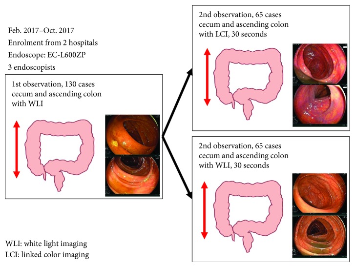
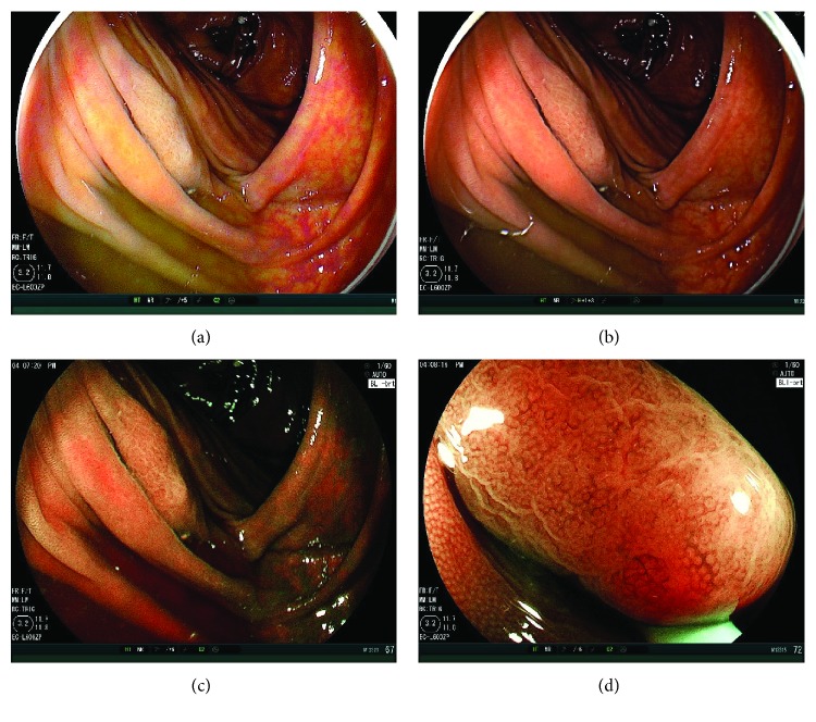
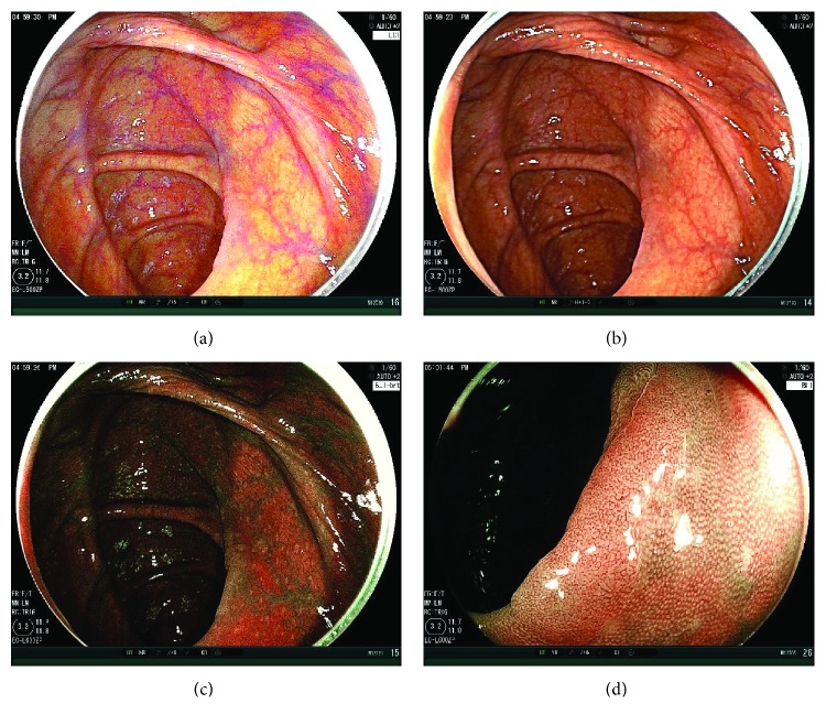
We enrolled patients undergoing colonoscopy from February to October 2017 in two institutions. In all patients, the cecum and ascending colon were observed with white light imaging (WLI) first. The colonoscope was inserted again, and the cecum and ascending colon were observed for an additional 30 seconds using either LCI or WLI. The method for the 30 sec observation was to insufflate the cecum and ascending colon sufficiently and observe them in a distant view, because the length of the second observation was determined to be precisely 30 sec. For the second observation, LCI was performed for the first 65 patients and WLI for the next 65. Adenoma and SSA/P detection rate (ASDR) in the second observation were examined in both groups. According to a pilot study, the sample size was estimated 65.

RESULTS

In the first observation, ASDR were 30.7% in the LCI group and 32.2% in the WLI group ( = 0.85). For the second observation, 13 polyps were detected in the LCI group and 5 polyps in the WLI group ( = 0.04). Additionally, ASDR for the second observation were 18.5% and 6.1%, respectively ( = 0.03). There were no significant differences between the LCI and WLI groups with respect to morphology (ratio of polypoid) (38.5% versus 60.0%, = 0.52) and histology (ratio of adenoma) (92.3% versus 100.0%, = 0.91). Total adenoma and SSA/P number were 48 in the LCI group and 36 in the WLI group ( = 0.02).

CONCLUSION

The 30 seconds additional observation with LCI improved the detection of adenoma and SSA/P in the right-sided colon.

摘要

背景与目的

漏诊息肉是结肠镜检查的一个隐患。在本研究中,我们分析了使用联动成像(LCI)额外观察30秒对检测腺瘤和无蒂锯齿状腺瘤/息肉(SSA/P)的有效性。

材料与方法

我们纳入了2017年2月至10月在两家机构接受结肠镜检查的患者。所有患者首先用白光成像(WLI)观察盲肠和升结肠。再次插入结肠镜,使用LCI或WLI对盲肠和升结肠额外观察30秒。30秒观察的方法是充分向盲肠和升结肠充气并在远距离观察,因为第二次观察的时长确定为精确的30秒。对于第二次观察,前65例患者采用LCI,后65例采用WLI。两组均检查第二次观察中的腺瘤和SSA/P检出率(ASDR)。根据一项初步研究,样本量估计为65例。

结果

在第一次观察中,LCI组的ASDR为30.7%,WLI组为32.2%(P = 0.85)。对于第二次观察,LCI组检测到13个息肉,WLI组检测到5个息肉(P = 0.04)。此外,第二次观察的ASDR分别为18.5%和6.1%(P = 0.03)。LCI组和WLI组在形态学(息肉样比例)(38.5%对60.0%,P = 0.52)和组织学(腺瘤比例)(92.3%对100.0%,P = 0.91)方面无显著差异。LCI组腺瘤和SSA/P总数为48个,WLI组为36个(P = 0.02)。

结论

使用LCI额外观察30秒可提高右侧结肠腺瘤和SSA/P的检出率。

https://cdn.ncbi.nlm.nih.gov/pmc/blobs/dba9/6079528/8c07c003aa32/GRP2018-5059834.003.jpg
https://cdn.ncbi.nlm.nih.gov/pmc/blobs/dba9/6079528/6f99ed6f432f/GRP2018-5059834.001.jpg
https://cdn.ncbi.nlm.nih.gov/pmc/blobs/dba9/6079528/0efcc5261f37/GRP2018-5059834.002.jpg
https://cdn.ncbi.nlm.nih.gov/pmc/blobs/dba9/6079528/8c07c003aa32/GRP2018-5059834.003.jpg
https://cdn.ncbi.nlm.nih.gov/pmc/blobs/dba9/6079528/6f99ed6f432f/GRP2018-5059834.001.jpg
https://cdn.ncbi.nlm.nih.gov/pmc/blobs/dba9/6079528/0efcc5261f37/GRP2018-5059834.002.jpg
https://cdn.ncbi.nlm.nih.gov/pmc/blobs/dba9/6079528/8c07c003aa32/GRP2018-5059834.003.jpg

相似文献

1
Additional Thirty Seconds Observation with Linked Color Imaging Improves Detection of Missed Polyps in the Right-Sided Colon.使用联动成像额外观察30秒可提高右侧结肠漏诊息肉的检出率。
Gastroenterol Res Pract. 2018 Jul 8;2018:5059834. doi: 10.1155/2018/5059834. eCollection 2018.
2
An Additional 30-s Observation of the Right-Sided Colon with Narrow Band Imaging Decreases Missed Polyps: A Pilot Study.窄带成像技术对右侧结肠额外观察 30 秒可减少遗漏息肉:一项初步研究。
Dig Dis Sci. 2018 Dec;63(12):3457-3464. doi: 10.1007/s10620-018-5275-1. Epub 2018 Sep 19.
3
Linked color imaging reduces the miss rate of neoplastic lesions in the right colon: a randomized tandem colonoscopy study.联动彩色成像降低右半结肠癌性病变的漏诊率:一项随机串联结肠镜检查研究
Endoscopy. 2018 Apr;50(4):396-402. doi: 10.1055/a-0580-7405. Epub 2018 Mar 14.
4
Additional 30-second observation of the right-sided colon for missed polyp detection with linked color imaging compared with narrow band imaging.与窄带成像相比,使用联动彩色成像对右侧结肠进行额外30秒观察以检测漏诊息肉。
Endosc Int Open. 2024 Oct 10;12(10):E1092-E1101. doi: 10.1055/a-2399-7554. eCollection 2024 Oct.
5
Linked color imaging enhances endoscopic detection of sessile serrated adenoma/polyps.联动成像增强了内镜下对无蒂锯齿状腺瘤/息肉的检测。
Endosc Int Open. 2018 Mar;6(3):E322-E334. doi: 10.1055/s-0043-124469. Epub 2018 Mar 7.
6
Linked color imaging versus white light imaging colonoscopy for colorectal adenoma detection: A randomized controlled trial.链接颜色成像与白光成像结肠镜检查在结直肠腺瘤检测中的比较:一项随机对照试验。
J Gastroenterol Hepatol. 2021 Oct;36(10):2778-2784. doi: 10.1111/jgh.15539. Epub 2021 May 20.
7
Colon polyp detection using linked color imaging compared to white light imaging: Systematic review and meta-analysis.利用链接色成像检测结肠息肉与白光成像的比较:系统评价和荟萃分析。
Dig Endosc. 2020 Sep;32(6):874-881. doi: 10.1111/den.13613. Epub 2020 Jan 30.
8
Impact of linked-color imaging on colorectal adenoma detection.联合显色成像对结直肠腺瘤检测的影响。
Gastrointest Endosc. 2019 Nov;90(5):826-834. doi: 10.1016/j.gie.2019.06.045. Epub 2019 Jul 11.
9
Linked-Color Imaging Detects More Colorectal Adenoma and Serrated Lesions: An International Randomized Controlled Trial.链接色成像检测到更多结直肠腺瘤和锯齿状病变:一项国际随机对照试验。
Clin Gastroenterol Hepatol. 2023 Jun;21(6):1493-1502.e4. doi: 10.1016/j.cgh.2022.10.021. Epub 2022 Nov 1.
10
Comparison Between Linked Color Imaging and Blue Laser Imaging for Improving the Visibility of Flat Colorectal Polyps: A Multicenter Pilot Study.链接显色成像与蓝激光成像改善平坦型结直肠息肉可视性的比较:一项多中心初步研究。
Dig Dis Sci. 2020 Jul;65(7):2054-2062. doi: 10.1007/s10620-019-05930-x. Epub 2019 Nov 14.

引用本文的文献

1
The impact of linked color imaging on adenoma detection rate in colonoscopy: a systematic review and meta-analysis.联动式彩色成像对结肠镜检查中腺瘤检出率的影响:一项系统评价和荟萃分析。
Clin Endosc. 2025 Mar;58(2):225-239. doi: 10.5946/ce.2024.072. Epub 2024 Oct 24.
2
Additional 30-second observation of the right-sided colon for missed polyp detection with linked color imaging compared with narrow band imaging.与窄带成像相比,使用联动彩色成像对右侧结肠进行额外30秒观察以检测漏诊息肉。
Endosc Int Open. 2024 Oct 10;12(10):E1092-E1101. doi: 10.1055/a-2399-7554. eCollection 2024 Oct.
3
Linked-color imaging with or without artificial intelligence for adenoma detection: a randomized trial.

本文引用的文献

1
Linked color imaging (LCI), a novel image-enhanced endoscopy technology, emphasizes the color of early gastric cancer.联动成像(LCI)是一种新型的图像增强内镜技术,它突出了早期胃癌的颜色。
Endosc Int Open. 2017 Oct;5(10):E1005-E1013. doi: 10.1055/s-0043-117881. Epub 2017 Oct 10.
2
Linked color imaging improves the visibility of various featured colorectal polyps in an endoscopist's visibility and color difference value.联动成像技术可提高内镜医师视野中各类特征性大肠息肉的可见度以及色差数值。
Int J Colorectal Dis. 2017 Sep;32(9):1253-1260. doi: 10.1007/s00384-017-2855-z. Epub 2017 Jul 19.
3
Risk factors of missed colorectal lesions after colonoscopy.
采用或不采用人工智能的联合同色成像用于腺瘤检测:一项随机试验
Endoscopy. 2024 May;56(5):376-383. doi: 10.1055/a-2239-8145. Epub 2024 Jan 8.
4
Detection of colorectal adenomas with texture and color enhancement imaging: Multicenter observational study.利用纹理和色彩增强成像检测结直肠腺瘤:多中心观察性研究。
Dig Endosc. 2023 May;35(4):529-537. doi: 10.1111/den.14480. Epub 2022 Dec 20.
5
The efficacy of polyp detection and tumor characterization of blue laser imaging, blue light imaging, and linked color imaging with light-emitted diode (LED) and LASER endoscope.蓝色激光成像、蓝光成像以及发光二极管(LED)和激光内窥镜的联合彩色成像在息肉检测和肿瘤特征描述方面的功效。
Ann Transl Med. 2020 Mar;8(5):152. doi: 10.21037/atm.2020.02.01.
6
The efficacy of tumor characterization and tumor detectability of linked color imaging and blue laser imaging with an LED endoscope compared to a LASER endoscope.与激光内窥镜相比,联用彩色成像和蓝色激光成像的发光二极管内窥镜在肿瘤特征识别及肿瘤可检测性方面的效能。
Int J Colorectal Dis. 2020 May;35(5):815-825. doi: 10.1007/s00384-020-03532-y. Epub 2020 Feb 22.
7
Ileal intubation is not associated with higher detection rate of right-sided conventional adenomas and serrated polyps compared to cecal intubation after adjustment for overall adenoma detection rate.经总体腺瘤检出率校正后,与盲肠插管相比,回肠插管与右侧传统腺瘤和锯齿状息肉的检出率升高无关。
BMC Gastroenterol. 2019 Nov 15;19(1):190. doi: 10.1186/s12876-019-1111-0.
8
Blue Laser Imaging, Blue Light Imaging, and Linked Color Imaging for the Detection and Characterization of Colorectal Tumors.蓝激光成像、蓝光成像和链接彩色成像在结直肠肿瘤检测和特征分析中的应用。
Gut Liver. 2019 Mar 15;13(2):140-148. doi: 10.5009/gnl18276.
结肠镜检查后结直肠病变漏诊的危险因素。
Medicine (Baltimore). 2017 Jul;96(27):e7468. doi: 10.1097/MD.0000000000007468.
4
Miss rate of colorectal neoplastic polyps and risk factors for missed polyps in consecutive colonoscopies.连续结肠镜检查中结直肠肿瘤性息肉的漏诊率及息肉漏诊的危险因素。
Intest Res. 2017 Jul;15(3):411-418. doi: 10.5217/ir.2017.15.3.411. Epub 2017 Jun 12.
5
Linked color imaging improves the visibility of colorectal polyps: a video study.联动彩色成像提高结直肠息肉的可视性:一项视频研究。
Endosc Int Open. 2017 Jun;5(6):E518-E525. doi: 10.1055/s-0043-105495. Epub 2017 Jun 7.
6
Comparison of linked color imaging and white-light colonoscopy for detection of colorectal polyps: a multicenter, randomized, crossover trial.比较蓝激光成像和白光结肠镜检查在结直肠息肉检测中的应用:一项多中心、随机、交叉试验。
Gastrointest Endosc. 2017 Oct;86(4):724-730. doi: 10.1016/j.gie.2017.02.035. Epub 2017 Mar 9.
7
Linked-color imaging improves endoscopic visibility of colorectal nongranular flat lesions.连码显色成像提高结直肠无颗粒平坦病变的内镜可视性。
Gastrointest Endosc. 2017 Oct;86(4):692-697. doi: 10.1016/j.gie.2017.01.044. Epub 2017 Feb 11.
8
Detectability of colorectal neoplastic lesions using a novel endoscopic system with blue laser imaging: a multicenter randomized controlled trial.新型内镜蓝激光成像系统检测结直肠肿瘤性病变的多中心随机对照研究。
Gastrointest Endosc. 2017 Aug;86(2):386-394. doi: 10.1016/j.gie.2017.01.017. Epub 2017 Jan 29.
9
Safety and Efficacy of a Same-Day Low-Volume 1 L PEG Bowel Preparation in Colonoscopy for the Elderly People and People with Renal Dysfunction.同日低容量1L聚乙二醇肠道准备法在老年及肾功能不全患者结肠镜检查中的安全性和有效性
Dig Dis Sci. 2016 Nov;61(11):3229-3235. doi: 10.1007/s10620-016-4262-7. Epub 2016 Aug 3.
10
Next-generation narrow band imaging system for colonic polyp detection: a prospective multicenter randomized trial.用于结肠息肉检测的新一代窄带成像系统:一项前瞻性多中心随机试验。
Int J Colorectal Dis. 2015 Jul;30(7):947-54. doi: 10.1007/s00384-015-2230-x. Epub 2015 Apr 30.