Biotechnology Center, Center for Molecular and Cellular Bioengineering, Technische Universität Dresden, Dresden, Germany.
Biotechnology Center, Center for Molecular and Cellular Bioengineering, Technische Universität Dresden, Dresden, Germany.
Biophys J. 2018 Sep 4;115(5):911-923. doi: 10.1016/j.bpj.2018.07.027. Epub 2018 Aug 4.
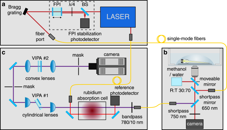
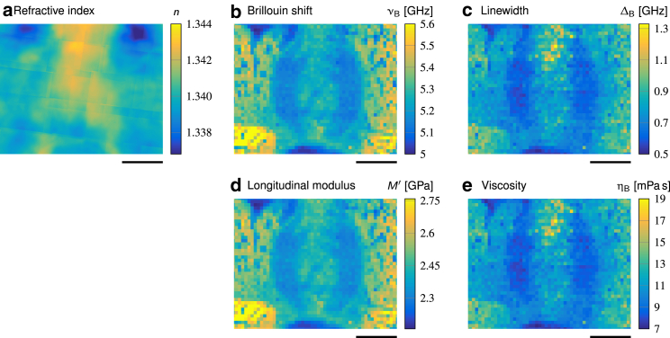
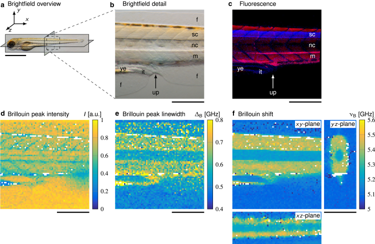
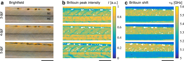
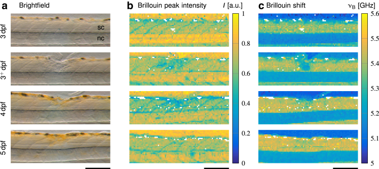
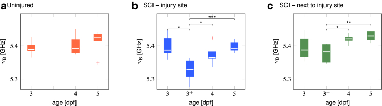
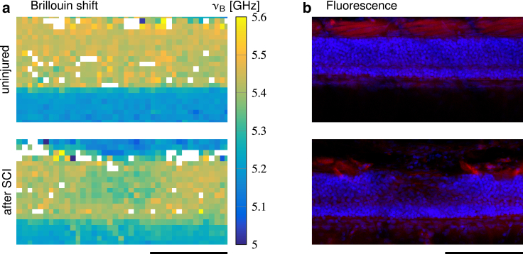
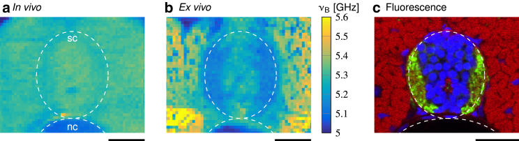
The mechanical properties of biological tissues are increasingly recognized as important factors in developmental and pathological processes. Most existing mechanical measurement techniques either necessitate destruction of the tissue for access or provide insufficient spatial resolution. Here, we show for the first time to our knowledge a systematic application of confocal Brillouin microscopy to quantitatively map the mechanical properties of spinal cord tissues during biologically relevant processes in a contact-free and nondestructive manner. Living zebrafish larvae were mechanically imaged in all anatomical planes during development and after spinal cord injury. These experiments revealed that Brillouin microscopy is capable of detecting the mechanical properties of distinct anatomical structures without interfering with the animal's natural development. The Brillouin shift within the spinal cord remained comparable during development and transiently decreased during the repair processes after spinal cord transection. By taking into account the refractive index distribution, we explicitly determined the apparent longitudinal modulus and viscosity of different larval zebrafish tissues. Importantly, mechanical properties differed between tissues in situ and in excised slices. The presented work constitutes the first step toward an in vivo assessment of spinal cord tissue mechanics during regeneration, provides a methodical basis to identify key determinants of mechanical tissue properties, and allows us to test their relative importance in combination with biochemical and genetic factors during developmental and regenerative processes.
生物组织的力学特性正逐渐被认为是发育和病理过程中的重要因素。大多数现有的力学测量技术要么需要破坏组织才能进行测量,要么提供的空间分辨率不足。在这里,我们首次在活体斑马鱼幼虫发育过程中和脊髓损伤后,以非接触和无损的方式,系统地应用共焦布里渊显微镜来定量绘制脊髓组织的力学特性。这些实验表明,布里渊显微镜能够在不干扰动物自然发育的情况下,检测到不同解剖结构的力学特性。在发育过程中,脊髓内的布里渊频移保持一致,而在脊髓横断后的修复过程中则短暂下降。通过考虑折射率分布,我们明确地确定了不同斑马鱼幼虫组织的表观纵弹性模量和黏度。重要的是,原位和离体组织的力学特性不同。本研究工作是在活体评估脊髓组织再生过程中的力学特性方面迈出的第一步,为确定力学组织特性的关键决定因素提供了方法学基础,并使我们能够在发育和再生过程中结合生化和遗传因素来测试它们的相对重要性。