• 文献检索
  • 文档翻译
  • 深度研究
  • 学术资讯
  • Suppr Zotero 插件Zotero 插件
  • 邀请有礼
  • 套餐&价格
  • 历史记录
应用&插件
Suppr Zotero 插件Zotero 插件浏览器插件Mac 客户端Windows 客户端微信小程序
定价
高级版会员购买积分包购买API积分包
服务
文献检索文档翻译深度研究API 文档MCP 服务
关于我们
关于 Suppr公司介绍联系我们用户协议隐私条款
关注我们

Suppr 超能文献

核心技术专利:CN118964589B侵权必究
粤ICP备2023148730 号-1Suppr @ 2026

文献检索

告别复杂PubMed语法,用中文像聊天一样搜索,搜遍4000万医学文献。AI智能推荐,让科研检索更轻松。

立即免费搜索

文件翻译

保留排版,准确专业,支持PDF/Word/PPT等文件格式,支持 12+语言互译。

免费翻译文档

深度研究

AI帮你快速写综述,25分钟生成高质量综述,智能提取关键信息,辅助科研写作。

立即免费体验

穹顶和锥体:ESCRT 蛋白诱导的膜黏附分裂。

Domes and cones: Adhesion-induced fission of membranes by ESCRT proteins.

机构信息

Theory & Bio-Systems Department, Max Planck Institute of Colloids and Interfaces, Potsdam, Germany.

Rudolf Peierls Centre for Theoretical Physics, University of Oxford, Oxford, United Kingdom.

出版信息

PLoS Comput Biol. 2018 Aug 21;14(8):e1006422. doi: 10.1371/journal.pcbi.1006422. eCollection 2018 Aug.

DOI:10.1371/journal.pcbi.1006422
PMID:30130367
原文链接:https://pmc.ncbi.nlm.nih.gov/articles/PMC6118396/
Abstract

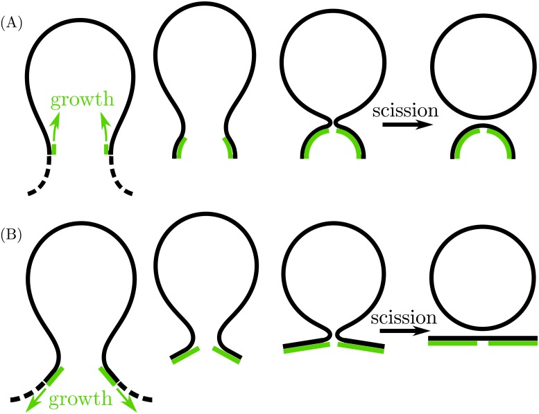
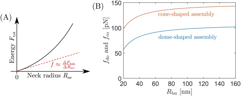
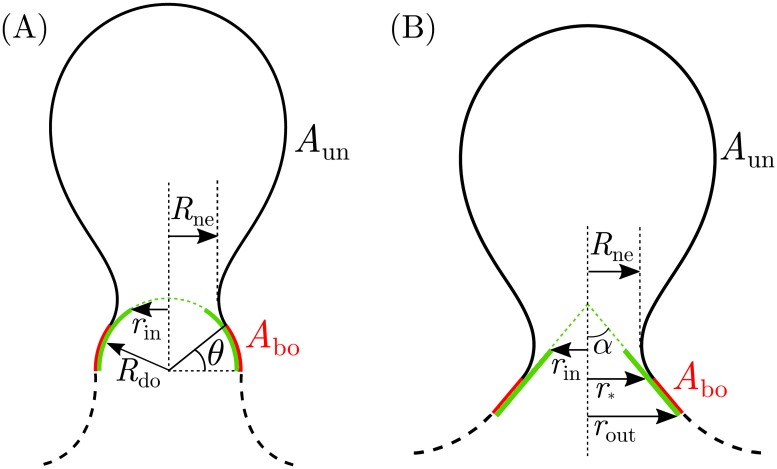
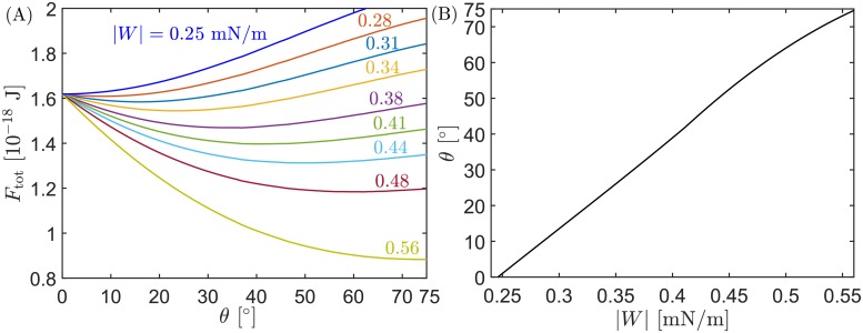
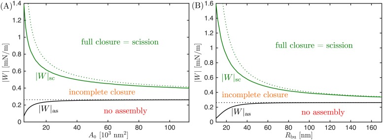
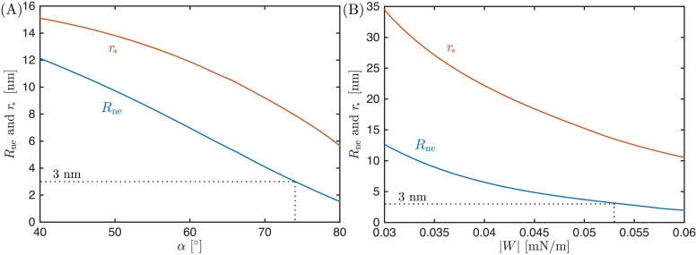
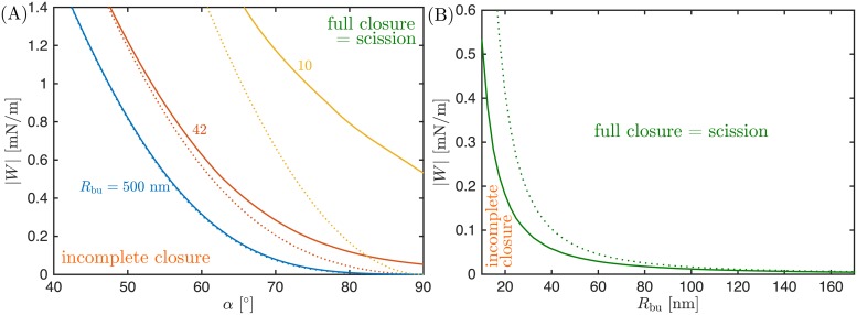
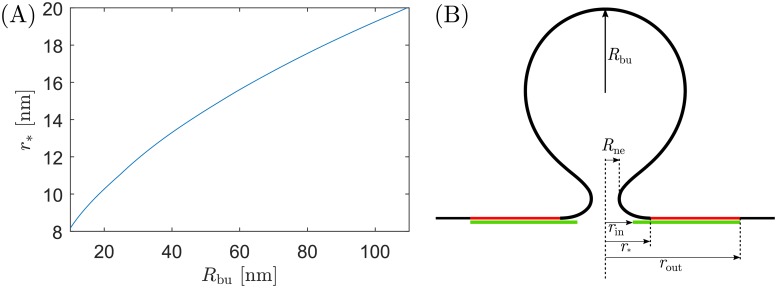
ESCRT proteins participate in the fission step of exocytic membrane budding, by assisting in the closure and scission of the membrane neck that connects the nascent bud to the plasma membrane. However, the precise mechanism by which the proteins achieve this so-called reverse-topology membrane scission remains to be elucidated. One mechanism is described by the dome model, which postulates that ESCRT-III proteins assemble in the shape of a hemispherical dome at the location of the neck, and guide the closure of this neck via membrane-protein adhesion. A different mechanism is described by the flattening cone model, in which the ESCRT-III complex first assembles at the neck in the shape of a cone, which then flattens leading to neck closure. Here, we use the theoretical framework of curvature elasticity and membrane-protein adhesion to quantitatively describe and compare both mechanisms. This comparison shows that the minimal adhesive strength of the membrane-protein interactions required for scission is much lower for cones than for domes, and that the geometric constraints on the shape of the assembly required to induce scission are more stringent for domes than for cones. Finally, we compute for the first time the adhesion-induced constriction forces exerted by the ESCRT assemblies onto the membrane necks. These forces are higher for cones and of the order of 100 pN.

摘要

ESCRT 蛋白参与胞吐膜出芽的裂变步骤,通过协助连接初生芽和质膜的膜颈的闭合和分裂。然而,这些蛋白质实现所谓的反向拓扑膜分裂的确切机制仍有待阐明。一种机制是由穹顶模型描述的,该模型假设 ESCRT-III 蛋白在颈部位置以半球形穹顶的形状组装,并通过膜蛋白粘附引导颈部的闭合。另一种机制是由扁平化锥体模型描述的,其中 ESCRT-III 复合物首先以锥体的形状在颈部组装,然后扁平化导致颈部闭合。在这里,我们使用曲率弹性和膜蛋白粘附的理论框架来定量描述和比较这两种机制。这种比较表明,对于锥体来说,用于分裂的膜蛋白相互作用的最小粘附强度要低得多,而对于穹顶来说,用于诱导分裂的组装形状的几何约束要比锥体更严格。最后,我们首次计算了 ESCRT 组装体施加在膜颈上的粘附诱导收缩力。对于锥体来说,这些力更高,约为 100 pN。

https://cdn.ncbi.nlm.nih.gov/pmc/blobs/744f/6118396/c3d176d9dcb0/pcbi.1006422.g008.jpg
https://cdn.ncbi.nlm.nih.gov/pmc/blobs/744f/6118396/bea91cf92f9c/pcbi.1006422.g001.jpg
https://cdn.ncbi.nlm.nih.gov/pmc/blobs/744f/6118396/a351222ff428/pcbi.1006422.g002.jpg
https://cdn.ncbi.nlm.nih.gov/pmc/blobs/744f/6118396/e2f0d07169ce/pcbi.1006422.g003.jpg
https://cdn.ncbi.nlm.nih.gov/pmc/blobs/744f/6118396/ca30f7336db3/pcbi.1006422.g004.jpg
https://cdn.ncbi.nlm.nih.gov/pmc/blobs/744f/6118396/5778caedb81d/pcbi.1006422.g005.jpg
https://cdn.ncbi.nlm.nih.gov/pmc/blobs/744f/6118396/23787383e4a4/pcbi.1006422.g006.jpg
https://cdn.ncbi.nlm.nih.gov/pmc/blobs/744f/6118396/cc0e134e45f3/pcbi.1006422.g007.jpg
https://cdn.ncbi.nlm.nih.gov/pmc/blobs/744f/6118396/c3d176d9dcb0/pcbi.1006422.g008.jpg

相似文献

1
Domes and cones: Adhesion-induced fission of membranes by ESCRT proteins.穹顶和锥体:ESCRT 蛋白诱导的膜黏附分裂。
PLoS Comput Biol. 2018 Aug 21;14(8):e1006422. doi: 10.1371/journal.pcbi.1006422. eCollection 2018 Aug.
2
Negative membrane curvature catalyzes nucleation of endosomal sorting complex required for transport (ESCRT)-III assembly.负膜曲率催化转运所需内体分选复合物(ESCRT)-III组装的成核过程。
Proc Natl Acad Sci U S A. 2015 Dec 29;112(52):15892-7. doi: 10.1073/pnas.1518765113. Epub 2015 Dec 14.
3
Assembly and disassembly of the ESCRT-III membrane scission complex.ESCRT-III 膜分裂复合物的组装和拆卸。
FEBS Lett. 2011 Oct 20;585(20):3191-6. doi: 10.1016/j.febslet.2011.09.001. Epub 2011 Sep 9.
4
ESCRT-III polymers in membrane neck constriction.ESCRT-III 聚合物在膜颈缢缩中的作用。
Trends Cell Biol. 2012 Mar;22(3):133-40. doi: 10.1016/j.tcb.2011.11.007. Epub 2012 Jan 10.
5
Recruitment dynamics of ESCRT-III and Vps4 to endosomes and implications for reverse membrane budding.内体上 ESCRT-III 和 Vps4 的募集动态及其对反向膜出芽的意义。
Elife. 2017 Oct 11;6:e31652. doi: 10.7554/eLife.31652.
6
Reverse-topology membrane scission by the ESCRT proteins.ESCRT蛋白介导的反向拓扑膜切割
Nat Rev Mol Cell Biol. 2017 Jan;18(1):5-17. doi: 10.1038/nrm.2016.121. Epub 2016 Oct 5.
7
Cellular Functions and Molecular Mechanisms of the ESCRT Membrane-Scission Machinery.ESCRT 膜分裂机器的细胞功能和分子机制。
Trends Biochem Sci. 2017 Jan;42(1):42-56. doi: 10.1016/j.tibs.2016.08.016. Epub 2016 Sep 23.
8
Computational model of membrane fission catalyzed by ESCRT-III.ESCRT-III 催化的膜裂变计算模型。
PLoS Comput Biol. 2009 Nov;5(11):e1000575. doi: 10.1371/journal.pcbi.1000575. Epub 2009 Nov 20.
9
The many functions of ESCRTs.ESCRTs 的多种功能。
Nat Rev Mol Cell Biol. 2020 Jan;21(1):25-42. doi: 10.1038/s41580-019-0177-4. Epub 2019 Nov 8.
10
Membrane budding and scission by the ESCRT machinery: it's all in the neck.通过 ESCRT 机制进行膜出芽和分裂:一切都在颈部。
Nat Rev Mol Cell Biol. 2010 Aug;11(8):556-66. doi: 10.1038/nrm2937. Epub 2010 Jun 30.

引用本文的文献

1
Dynamics of upstream ESCRT organization at the HIV-1 budding site.HIV-1 出芽位点处的上游 ESCRT 组织动力学。
Biophys J. 2023 Jul 11;122(13):2655-2674. doi: 10.1016/j.bpj.2023.05.020. Epub 2023 May 22.
2
Stepwise remodeling and subcompartment formation in individual vesicles by three ESCRT-III proteins.三种ESCRT-III蛋白在单个囊泡中进行逐步重塑和亚区室形成。
iScience. 2022 Dec 8;26(1):105765. doi: 10.1016/j.isci.2022.105765. eCollection 2023 Jan 20.
3
Snf7 spirals sense and alter membrane curvature.Snf7 螺旋感知并改变膜曲率。

本文引用的文献

1
The Conserved ESCRT-III Machinery Participates in the Phagocytosis of .保守的 ESCRT-III 机器参与. 的吞噬作用。
Front Cell Infect Microbiol. 2018 Mar 1;8:53. doi: 10.3389/fcimb.2018.00053. eCollection 2018.
2
Recruitment dynamics of ESCRT-III and Vps4 to endosomes and implications for reverse membrane budding.内体上 ESCRT-III 和 Vps4 的募集动态及其对反向膜出芽的意义。
Elife. 2017 Oct 11;6:e31652. doi: 10.7554/eLife.31652.
3
Dynamic subunit turnover in ESCRT-III assemblies is regulated by Vps4 to mediate membrane remodelling during cytokinesis.
Nat Commun. 2022 Apr 21;13(1):2174. doi: 10.1038/s41467-022-29850-z.
4
Protein crowding mediates membrane remodeling in upstream ESCRT-induced formation of intraluminal vesicles.蛋白质拥挤介导了在上游 ESCRT 诱导的腔内小泡形成过程中的膜重塑。
Proc Natl Acad Sci U S A. 2020 Nov 17;117(46):28614-28624. doi: 10.1073/pnas.2014228117. Epub 2020 Nov 2.
5
The ESCRTs - converging on mechanism.ESCRTs-汇聚于机制。
J Cell Sci. 2020 Sep 16;133(18):jcs240333. doi: 10.1242/jcs.240333.
6
Biophysical forces in membrane bending and traffic.膜弯曲和运输中的生物物理力。
Curr Opin Cell Biol. 2020 Aug;65:72-77. doi: 10.1016/j.ceb.2020.02.017. Epub 2020 Mar 28.
7
Molecular Simulation of Mechanical Properties and Membrane Activities of the ESCRT-III Complexes.ESCRT-III复合物力学性质与膜活性的分子模拟
Biophys J. 2020 Mar 24;118(6):1333-1343. doi: 10.1016/j.bpj.2020.01.033. Epub 2020 Feb 4.
8
Changes in ESCRT-III filament geometry drive membrane remodelling and fission in silico.ESCRT-III 丝状体几何形状的变化在计算机中驱动膜重塑和裂变。
BMC Biol. 2019 Oct 22;17(1):82. doi: 10.1186/s12915-019-0700-2.
9
In Vitro Membrane Remodeling by ESCRT is Regulated by Negative Feedback from Membrane Tension.ESCRT介导的体外膜重塑受膜张力负反馈调节。
iScience. 2019 May 31;15:173-184. doi: 10.1016/j.isci.2019.04.021. Epub 2019 Apr 20.
10
Modeling Membrane Curvature Generation due to Membrane⁻Protein Interactions.模拟膜蛋白相互作用引起的膜曲率生成。
Biomolecules. 2018 Oct 23;8(4):120. doi: 10.3390/biom8040120.
ESCRT-III组装体中的动态亚基周转由Vps4调节,以在胞质分裂期间介导膜重塑。
Nat Cell Biol. 2017 Jul;19(7):787-798. doi: 10.1038/ncb3559. Epub 2017 Jun 12.
4
LEM2 recruits CHMP7 for ESCRT-mediated nuclear envelope closure in fission yeast and human cells.在裂殖酵母和人类细胞中,LEM2招募CHMP7以进行ESCRT介导的核膜封闭。
Proc Natl Acad Sci U S A. 2017 Mar 14;114(11):E2166-E2175. doi: 10.1073/pnas.1613916114. Epub 2017 Feb 27.
5
Uniform and Janus-like nanoparticles in contact with vesicles: energy landscapes and curvature-induced forces.具有各向同性和双面性的纳米粒子与囊泡接触:能量景观和曲率诱导力。
Soft Matter. 2017 Mar 15;13(11):2155-2173. doi: 10.1039/c6sm02796b.
6
Reverse-topology membrane scission by the ESCRT proteins.ESCRT蛋白介导的反向拓扑膜切割
Nat Rev Mol Cell Biol. 2017 Jan;18(1):5-17. doi: 10.1038/nrm.2016.121. Epub 2016 Oct 5.
7
Stabilization of membrane necks by adhesive particles, substrate surfaces, and constriction forces.通过粘性颗粒、基底表面和压缩力稳定膜颈部。
Soft Matter. 2016 Oct 21;12(39):8155-8166. doi: 10.1039/c6sm01481j. Epub 2016 Aug 10.
8
Structure and membrane remodeling activity of ESCRT-III helical polymers.内体分选转运复合体Ⅲ(ESCRT-III)螺旋聚合物的结构与膜重塑活性
Science. 2015 Dec 18;350(6267):1548-51. doi: 10.1126/science.aad8305. Epub 2015 Dec 3.
9
ESCRT Filaments as Spiral Springs.ESCRT 丝作为螺旋弹簧。
Dev Cell. 2015 Nov 23;35(4):397-8. doi: 10.1016/j.devcel.2015.11.007.
10
Relaxation of Loaded ESCRT-III Spiral Springs Drives Membrane Deformation.负载的ESCRT-III螺旋弹簧的松弛驱动膜变形。
Cell. 2015 Nov 5;163(4):866-79. doi: 10.1016/j.cell.2015.10.017. Epub 2015 Oct 29.