Department of Radiology and Medical Imaging, University of Virginia, Charlottesville, Virginia.
Cingulate, Hampton, New Hampshire.
Acad Radiol. 2019 Mar;26(3):412-423. doi: 10.1016/j.acra.2018.08.003. Epub 2018 Sep 6.
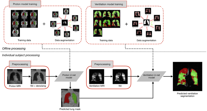
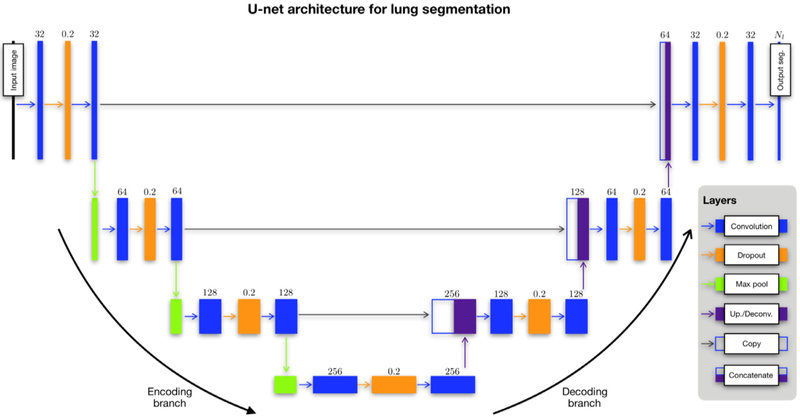
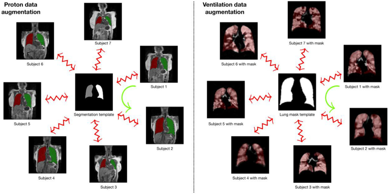
We propose an automated segmentation pipeline based on deep learning for proton lung MRI segmentation and ventilation-based quantification which improves on our previously reported methodologies in terms of computational efficiency while demonstrating accuracy and robustness. The large data requirement for the proposed framework is made possible by a novel template-based data augmentation strategy. Supporting this work is the open-source ANTsRNet-a growing repository of well-known deep learning architectures first introduced here.
Deep convolutional neural network (CNN) models were constructed and trained using a custom multilabel Dice metric loss function and a novel template-based data augmentation strategy. Training (including template generation and data augmentation) employed 205 proton MR images and 73 functional lung MRI. Evaluation was performed using data sets of size 63 and 40 images, respectively.
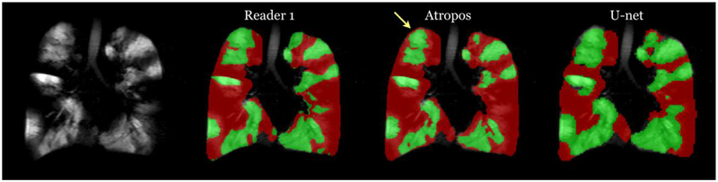
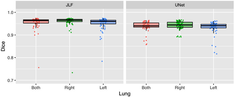
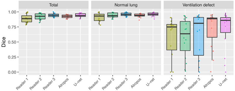
Accuracy for CNN-based proton lung MRI segmentation (in terms of Dice overlap) was left lung: 0.93 ± 0.03, right lung: 0.94 ± 0.02, and whole lung: 0.94 ± 0.02. Although slightly less accurate than our previously reported joint label fusion approach (left lung: 0.95 ± 0.02, right lung: 0.96 ± 0.01, and whole lung: 0.96 ± 0.01), processing time is <1 second per subject for the proposed approach versus ∼30 minutes per subject using joint label fusion. Accuracy for quantifying ventilation defects was determined based on a consensus labeling where average accuracy (Dice multilabel overlap of ventilation defect regions plus normal region) was 0.94 for the CNN method; 0.92 for our previously reported method; and 0.90, 0.92, and 0.94 for expert readers.
The proposed framework yields accurate automated quantification in near real time. CNNs drastically reduce processing time after offline model construction and demonstrate significant future potential for facilitating quantitative analysis of functional lung MRI.
我们提出了一种基于深度学习的质子肺部 MRI 分割和通气定量的自动分割管道,在计算效率方面优于我们之前报道的方法,同时展示了准确性和鲁棒性。所提出框架的大数据需求得益于一种新颖的基于模板的数据扩充策略。支持这项工作的是开源的 ANTsRNet——一个在这里首次引入的知名深度学习架构的不断增长的存储库。
使用定制的多标签 Dice 度量损失函数和新颖的基于模板的数据扩充策略构建和训练深度卷积神经网络 (CNN) 模型。训练(包括模板生成和数据扩充)使用了 205 个质子 MRI 图像和 73 个功能肺部 MRI。评估分别使用大小为 63 和 40 张图像的数据集进行。
基于 CNN 的质子肺部 MRI 分割的准确性(以 Dice 重叠表示)为左肺:0.93 ± 0.03,右肺:0.94 ± 0.02,全肺:0.94 ± 0.02。尽管略低于我们之前报道的联合标签融合方法(左肺:0.95 ± 0.02,右肺:0.96 ± 0.01,全肺:0.96 ± 0.01),但对于所提出的方法,每个患者的处理时间不到 1 秒,而使用联合标签融合方法则为每个患者约 30 分钟。通气缺陷定量的准确性是基于共识标记确定的,其中 CNN 方法的平均准确性(通气缺陷区域和正常区域的多标签 Dice 重叠)为 0.94;我们之前报道的方法为 0.92;专家读者的准确性分别为 0.90、0.92 和 0.94。
所提出的框架可在近实时情况下实现准确的自动定量。在离线模型构建后,CNN 大大减少了处理时间,并为促进功能肺部 MRI 的定量分析展示了巨大的未来潜力。