• 文献检索
  • 文档翻译
  • 深度研究
  • 学术资讯
  • Suppr Zotero 插件Zotero 插件
  • 邀请有礼
  • 套餐&价格
  • 历史记录
应用&插件
Suppr Zotero 插件Zotero 插件浏览器插件Mac 客户端Windows 客户端微信小程序
定价
高级版会员购买积分包购买API积分包
服务
文献检索文档翻译深度研究API 文档MCP 服务
关于我们
关于 Suppr公司介绍联系我们用户协议隐私条款
关注我们

Suppr 超能文献

核心技术专利:CN118964589B侵权必究
粤ICP备2023148730 号-1Suppr @ 2026

文献检索

告别复杂PubMed语法,用中文像聊天一样搜索,搜遍4000万医学文献。AI智能推荐,让科研检索更轻松。

立即免费搜索

文件翻译

保留排版,准确专业,支持PDF/Word/PPT等文件格式,支持 12+语言互译。

免费翻译文档

深度研究

AI帮你快速写综述,25分钟生成高质量综述,智能提取关键信息,辅助科研写作。

立即免费体验

通过完整颅骨对小鼠大脑结构和功能进行三光子成像。

Three-photon imaging of mouse brain structure and function through the intact skull.

机构信息

School of Applied and Engineering Physics, Cornell University, Ithaca, NY, USA.

CNC Program, Stanford University, Stanford, CA, USA.

出版信息

Nat Methods. 2018 Oct;15(10):789-792. doi: 10.1038/s41592-018-0115-y. Epub 2018 Sep 10.

DOI:10.1038/s41592-018-0115-y
PMID:30202059
原文链接:https://pmc.ncbi.nlm.nih.gov/articles/PMC6188644/
Abstract

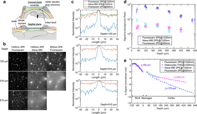
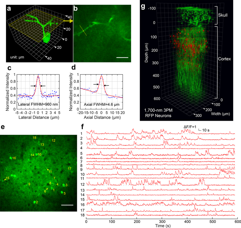
Optical imaging through the intact mouse skull is challenging because of skull-induced aberrations and scattering. We found that three-photon excitation provided improved optical sectioning compared with that obtained with two-photon excitation, even when we used the same excitation wavelength and imaging system. Here we demonstrate three-photon imaging of vasculature through the adult mouse skull at >500-μm depth, as well as GCaMP6s calcium imaging over weeks in cortical layers 2/3 and 4 in awake mice, with 8.5 frames per second and a field of view spanning hundreds of micrometers.

摘要

通过完整的小鼠颅骨进行光学成像是具有挑战性的,因为颅骨会引起像差和散射。我们发现,与双光子激发相比,三光子激发提供了更好的光学切片,即使我们使用相同的激发波长和成像系统。在这里,我们展示了通过成年小鼠颅骨进行的三光子血管成像,深度超过 500μm,以及在清醒小鼠的皮层 2/3 和 4 层中进行的 GCaMP6s 钙成像,帧率为每秒 8.5 帧,视场跨越数百微米。

https://cdn.ncbi.nlm.nih.gov/pmc/blobs/9d35/6188644/a1ec33dc5006/nihms-1502265-f0002.jpg
https://cdn.ncbi.nlm.nih.gov/pmc/blobs/9d35/6188644/fccf986f0262/nihms-1502265-f0001.jpg
https://cdn.ncbi.nlm.nih.gov/pmc/blobs/9d35/6188644/a1ec33dc5006/nihms-1502265-f0002.jpg
https://cdn.ncbi.nlm.nih.gov/pmc/blobs/9d35/6188644/fccf986f0262/nihms-1502265-f0001.jpg
https://cdn.ncbi.nlm.nih.gov/pmc/blobs/9d35/6188644/a1ec33dc5006/nihms-1502265-f0002.jpg

相似文献

1
Three-photon imaging of mouse brain structure and function through the intact skull.通过完整颅骨对小鼠大脑结构和功能进行三光子成像。
Nat Methods. 2018 Oct;15(10):789-792. doi: 10.1038/s41592-018-0115-y. Epub 2018 Sep 10.
2
High-resolution in vivo imaging of mouse brain through the intact skull.通过完整颅骨对小鼠大脑进行高分辨率活体成像。
Proc Natl Acad Sci U S A. 2015 Jul 28;112(30):9236-41. doi: 10.1073/pnas.1505939112. Epub 2015 Jul 13.
3
High-resolution structural and functional deep brain imaging using adaptive optics three-photon microscopy.基于自适应光学的三光子显微镜的高分辨率结构和功能深脑成像。
Nat Methods. 2021 Oct;18(10):1253-1258. doi: 10.1038/s41592-021-01257-6. Epub 2021 Sep 30.
4
In vivo three-photon imaging of activity of GCaMP6-labeled neurons deep in intact mouse brain.在完整小鼠大脑深处用 GCaMP6 标记的神经元的体内三光子成像。
Nat Methods. 2017 Apr;14(4):388-390. doi: 10.1038/nmeth.4183. Epub 2017 Feb 20.
5
Miniature two-photon microscopy for enlarged field-of-view, multi-plane and long-term brain imaging.用于大视场、多平面和长期脑成像的微型双光子显微镜。
Nat Methods. 2021 Jan;18(1):46-49. doi: 10.1038/s41592-020-01024-z. Epub 2021 Jan 6.
6
Three-photon head-mounted microscope for imaging deep cortical layers in freely moving rats.三光子头佩戴显微镜,用于在自由活动的大鼠中成像深层皮层。
Nat Methods. 2020 May;17(5):509-513. doi: 10.1038/s41592-020-0817-9. Epub 2020 May 4.
7
Facile Synthesis of Efficient Luminogens with AIE Features for Three-Photon Fluorescence Imaging of the Brain through the Intact Skull.通过完整颅骨的三光子荧光成像对大脑进行高效发光体的简便合成,具有 AIE 特性。
Adv Mater. 2020 Jun;32(23):e2000364. doi: 10.1002/adma.202000364. Epub 2020 Apr 30.
8
Transcranial two-photon imaging of the living mouse brain.活体小鼠大脑的经颅双光子成像。
Cold Spring Harb Protoc. 2011 Sep 1;2011(9):10.1101/pdb.prot065474 pdb.prot065474. doi: 10.1101/pdb.prot065474.
9
Improved deep two-photon calcium imaging in vivo.体内深度双光子钙成像的改进
Cell Calcium. 2017 Jun;64:29-35. doi: 10.1016/j.ceca.2016.12.005. Epub 2016 Dec 21.
10
All-near-infrared multiphoton microscopy interrogates intact tissues at deeper imaging depths than conventional single- and two-photon near-infrared excitation microscopes.全近红外多光子显微镜比传统的单光子和双光子近红外激发显微镜能够在更深的成像深度下对完整组织进行探测。
J Biomed Opt. 2013 Oct;18(10):106012. doi: 10.1117/1.JBO.18.10.106012.

引用本文的文献

1
Advances in two-photon imaging for monitoring neural activity in behaving mice.用于监测行为小鼠神经活动的双光子成像技术进展。
Front Neurosci. 2025 Aug 11;19:1597151. doi: 10.3389/fnins.2025.1597151. eCollection 2025.
2
SeeThrough: a rationally designed skull clearing technique for in vivo brain imaging.SeeThrough:一种用于体内脑成像的合理设计的颅骨清除技术。
Nat Commun. 2025 Aug 26;16(1):7584. doi: 10.1038/s41467-025-62836-1.
3
Photobleaching Analysis of Fluorescent Proteins in Two-Photon Microscopy at High Repetition Rates.

本文引用的文献

1
Skull optical clearing window for imaging of the mouse cortex at synaptic resolution.用于在突触分辨率下对小鼠皮层进行成像的颅骨光学透明窗口。
Light Sci Appl. 2018 Feb 23;7:17153. doi: 10.1038/lsa.2017.153. eCollection 2018.
2
A large, switchable optical clearing skull window for cerebrovascular imaging.一种用于脑血管成像的大型、可切换光透明颅骨窗口。
Theranostics. 2018 Apr 9;8(10):2696-2708. doi: 10.7150/thno.23686. eCollection 2018.
3
Aggregation-Induced Emission Luminogen with Deep-Red Emission for Through-Skull Three-Photon Fluorescence Imaging of Mouse.
高重复率双光子显微镜下荧光蛋白的光漂白分析
Opt Commun. 2025 Jan 1;574. doi: 10.1016/j.optcom.2024.131059. Epub 2024 Sep 3.
4
Compressive Fourier-Domain Intensity Coupling (C-FOCUS) enables near-millimeter deep imaging in the intact mouse brain in vivo.压缩傅里叶域强度耦合(C-FOCUS)能够在完整的活体小鼠大脑中实现近毫米深度成像。
Res Sq. 2025 Jul 28:rs.3.rs-7104566. doi: 10.21203/rs.3.rs-7104566/v1.
5
Multiphoton Neurophotonics: Recent Advances in Imaging and Manipulating Neuronal Circuits.多光子神经光子学:成像与操纵神经回路的最新进展
ACS Photonics. 2025 Apr 4;12(7):3296-3318. doi: 10.1021/acsphotonics.4c02101. eCollection 2025 Jul 16.
6
Three-photon microscopy: an emerging technique for deep intravital brain imaging.三光子显微镜:一种用于深层活体脑成像的新兴技术。
Nat Rev Neurosci. 2025 Jun 20. doi: 10.1038/s41583-025-00937-y.
7
Three-photon in vivo imaging of neurons and glia in the medial prefrontal cortex with sub-cellular resolution.内侧前额叶皮质中神经元和神经胶质细胞的三光子亚细胞分辨率体内成像。
Commun Biol. 2025 May 23;8(1):795. doi: 10.1038/s42003-025-08079-8.
8
Protocol to analyze marrow B lineage cell dynamics by in vivo three-photon microscopy in intact mouse tibia.通过体内三光子显微镜分析完整小鼠胫骨中骨髓B淋巴细胞动态变化的实验方案。
STAR Protoc. 2025 May 14;6(2):103824. doi: 10.1016/j.xpro.2025.103824.
9
Reconstructing Three-Dimensional Optical Anisotropy with Tomographic Müller-Polarimetric Microscopy.用层析穆勒偏振显微镜重建三维光学各向异性
Adv Sci (Weinh). 2025 Jul;12(27):e2502075. doi: 10.1002/advs.202502075. Epub 2025 May 8.
10
Color-neutral and reversible tissue transparency enables longitudinal deep-tissue imaging in live mice.颜色中性且可逆的组织透明化能够实现对活体小鼠进行纵向深度组织成像。
bioRxiv. 2025 Feb 27:2025.02.20.639185. doi: 10.1101/2025.02.20.639185.
用于小鼠颅透过三光子荧光成像的深红光发射聚集诱导发光团。
ACS Nano. 2017 Oct 24;11(10):10452-10461. doi: 10.1021/acsnano.7b05645. Epub 2017 Oct 12.
4
Chronic imaging through "transparent skull" in mice.通过“透明颅骨”对小鼠进行长期成像。
PLoS One. 2017 Aug 16;12(8):e0181788. doi: 10.1371/journal.pone.0181788. eCollection 2017.
5
In vivo three-photon imaging of activity of GCaMP6-labeled neurons deep in intact mouse brain.在完整小鼠大脑深处用 GCaMP6 标记的神经元的体内三光子成像。
Nat Methods. 2017 Apr;14(4):388-390. doi: 10.1038/nmeth.4183. Epub 2017 Feb 20.
6
Through-skull fluorescence imaging of the brain in a new near-infrared window.在一个新的近红外窗口对大脑进行经颅荧光成像。
Nat Photonics. 2014 Sep;8(9):723-730. doi: 10.1038/nphoton.2014.166. Epub 2014 Aug 3.
7
Brain heating induced by near-infrared lasers during multiphoton microscopy.多光子显微镜检查期间近红外激光引起的脑部发热
J Neurophysiol. 2016 Sep 1;116(3):1012-23. doi: 10.1152/jn.00275.2016. Epub 2016 Jun 8.
8
Intact skull chronic windows for mesoscopic wide-field imaging in awake mice.用于清醒小鼠介观宽视场成像的完整颅骨慢性窗口
J Neurosci Methods. 2016 Jul 15;267:141-9. doi: 10.1016/j.jneumeth.2016.04.012. Epub 2016 Apr 19.
9
In vivo Calcium Imaging of Evoked Calcium Waves in the Embryonic Cortex.胚胎皮质中诱发钙波的体内钙成像
Front Cell Neurosci. 2016 Jan 6;9:500. doi: 10.3389/fncel.2015.00500. eCollection 2015.
10
The power of using functional fMRI on small rodents to study brain pharmacology and disease.在小型啮齿动物身上使用功能磁共振成像来研究脑药理学和疾病的作用。
Front Pharmacol. 2015 Oct 21;6:231. doi: 10.3389/fphar.2015.00231. eCollection 2015.