• 文献检索
  • 文档翻译
  • 深度研究
  • 学术资讯
  • Suppr Zotero 插件Zotero 插件
  • 邀请有礼
  • 套餐&价格
  • 历史记录
应用&插件
Suppr Zotero 插件Zotero 插件浏览器插件Mac 客户端Windows 客户端微信小程序
定价
高级版会员购买积分包购买API积分包
服务
文献检索文档翻译深度研究API 文档MCP 服务
关于我们
关于 Suppr公司介绍联系我们用户协议隐私条款
关注我们

Suppr 超能文献

核心技术专利:CN118964589B侵权必究
粤ICP备2023148730 号-1Suppr @ 2026

文献检索

告别复杂PubMed语法,用中文像聊天一样搜索,搜遍4000万医学文献。AI智能推荐,让科研检索更轻松。

立即免费搜索

文件翻译

保留排版,准确专业,支持PDF/Word/PPT等文件格式,支持 12+语言互译。

免费翻译文档

深度研究

AI帮你快速写综述,25分钟生成高质量综述,智能提取关键信息,辅助科研写作。

立即免费体验

脊柱结构的可塑性:局部信号传导、翻译与细胞骨架重组

Plasticity of Spine Structure: Local Signaling, Translation and Cytoskeletal Reorganization.

作者信息

Nakahata Yoshihisa, Yasuda Ryohei

机构信息

Neuronal Signal Transduction, Max Planck Florida Institute for Neuroscience (MPFI), Jupiter, FL, United States.

出版信息

Front Synaptic Neurosci. 2018 Aug 29;10:29. doi: 10.3389/fnsyn.2018.00029. eCollection 2018.

DOI:10.3389/fnsyn.2018.00029
PMID:30210329
原文链接:https://pmc.ncbi.nlm.nih.gov/articles/PMC6123351/
Abstract

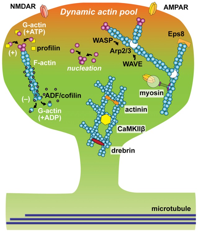
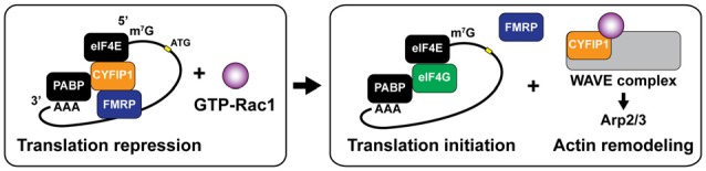
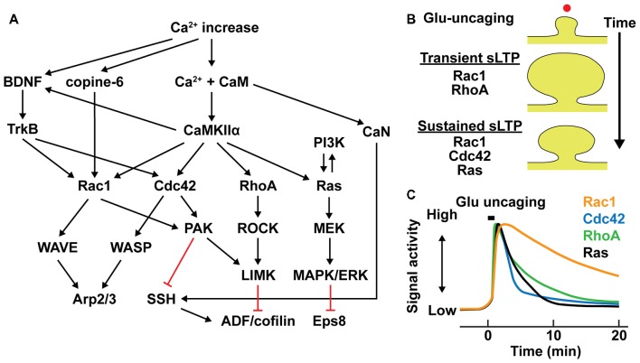
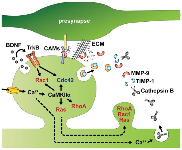
Dendritic spines are small protrusive structures on dendritic surfaces, and function as postsynaptic compartments for excitatory synapses. Plasticity of spine structure is associated with many forms of long-term neuronal plasticity, learning and memory. Inside these small dendritic compartments, biochemical states and protein-protein interactions are dynamically modulated by synaptic activity, leading to the regulation of protein synthesis and reorganization of cytoskeletal architecture. This in turn causes plasticity of structure and function of the spine. Technical advances in monitoring molecular behaviors in single dendritic spines have revealed that each signaling pathway is differently regulated across multiple spatiotemporal domains. The spatial pattern of signaling activity expands from a single spine to the nearby dendritic area, dendritic branch and the nucleus, regulating different cellular events at each spatial scale. Temporally, biochemical events are typically triggered by short Ca pulses (~10-100 ms). However, these signals can then trigger activation of downstream protein cascades that can last from milliseconds to hours. Recent imaging studies provide many insights into the biochemical processes governing signaling events of molecular assemblies at different spatial localizations. Here, we highlight recent findings of signaling dynamics during synaptic plasticity and discuss their roles in long-term structural plasticity of dendritic spines.

摘要

树突棘是树突表面的小突起结构,作为兴奋性突触的突触后区室发挥作用。树突棘结构的可塑性与多种形式的长期神经元可塑性、学习和记忆相关。在这些小的树突区室内,生化状态和蛋白质 - 蛋白质相互作用受到突触活动的动态调节,从而导致蛋白质合成的调节和细胞骨架结构的重组。这进而引起树突棘结构和功能的可塑性。监测单个树突棘中分子行为的技术进步表明,每个信号通路在多个时空域中受到不同的调节。信号活动的空间模式从单个树突棘扩展到附近的树突区域、树突分支和细胞核,在每个空间尺度上调节不同的细胞事件。在时间上,生化事件通常由短暂的钙脉冲(约10 - 100毫秒)触发。然而,这些信号随后可以触发下游蛋白质级联反应的激活,其持续时间可以从毫秒到数小时。最近的成像研究为不同空间定位的分子组装信号事件的生化过程提供了许多见解。在这里,我们重点介绍突触可塑性期间信号动力学的最新发现,并讨论它们在树突棘长期结构可塑性中的作用。

https://cdn.ncbi.nlm.nih.gov/pmc/blobs/b2d4/6123351/752fa800925a/fnsyn-10-00029-g0005.jpg
https://cdn.ncbi.nlm.nih.gov/pmc/blobs/b2d4/6123351/fc1f87add4f3/fnsyn-10-00029-g0001.jpg
https://cdn.ncbi.nlm.nih.gov/pmc/blobs/b2d4/6123351/27e09c81956f/fnsyn-10-00029-g0002.jpg
https://cdn.ncbi.nlm.nih.gov/pmc/blobs/b2d4/6123351/04c95f4b4488/fnsyn-10-00029-g0003.jpg
https://cdn.ncbi.nlm.nih.gov/pmc/blobs/b2d4/6123351/ad8a0e9f349f/fnsyn-10-00029-g0004.jpg
https://cdn.ncbi.nlm.nih.gov/pmc/blobs/b2d4/6123351/752fa800925a/fnsyn-10-00029-g0005.jpg

相似文献

1
Plasticity of Spine Structure: Local Signaling, Translation and Cytoskeletal Reorganization.脊柱结构的可塑性:局部信号传导、翻译与细胞骨架重组
Front Synaptic Neurosci. 2018 Aug 29;10:29. doi: 10.3389/fnsyn.2018.00029. eCollection 2018.
2
Plasticity of dendritic spines: Molecular function and dysfunction in neurodevelopmental disorders.树突棘可塑性:神经发育障碍中的分子功能与功能障碍。
Psychiatry Clin Neurosci. 2019 Sep;73(9):541-550. doi: 10.1111/pcn.12899. Epub 2019 Jul 8.
3
Actin Tyrosine-53-Phosphorylation in Neuronal Maturation and Synaptic Plasticity.神经元成熟和突触可塑性过程中的肌动蛋白酪氨酸-53磷酸化
J Neurosci. 2016 May 11;36(19):5299-313. doi: 10.1523/JNEUROSCI.2649-15.2016.
4
Imaging dendritic spines: molecular organization and signaling for plasticity.成像树突棘:可塑性的分子组织与信号传导
Curr Opin Neurobiol. 2021 Apr;67:66-74. doi: 10.1016/j.conb.2020.08.006. Epub 2020 Sep 15.
5
Biophysics of Biochemical Signaling in Dendritic Spines: Implications in Synaptic Plasticity.树突棘中生化信号的生物物理学:对突触可塑性的影响
Biophys J. 2017 Nov 21;113(10):2152-2159. doi: 10.1016/j.bpj.2017.07.029. Epub 2017 Aug 30.
6
Regulation of neuronal PKA signaling through AKAP targeting dynamics.通过A激酶锚定蛋白靶向动力学对神经元蛋白激酶A信号传导的调节
Eur J Cell Biol. 2006 Jul;85(7):627-33. doi: 10.1016/j.ejcb.2006.01.010. Epub 2006 Feb 28.
7
Postsynaptic signaling during plasticity of dendritic spines.树突棘可塑性过程中的突触后信号传递。
Trends Neurosci. 2012 Feb;35(2):135-43. doi: 10.1016/j.tins.2011.12.002. Epub 2012 Jan 3.
8
The structure and function of actin cytoskeleton in mature glutamatergic dendritic spines.成熟谷氨酸能树突棘中肌动蛋白细胞骨架的结构与功能。
Brain Res. 2014 Jul 21;1573:1-16. doi: 10.1016/j.brainres.2014.05.024. Epub 2014 May 20.
9
Dendritic spines: Revisiting the physiological role.树突棘:重新审视其生理作用。
Prog Neuropsychopharmacol Biol Psychiatry. 2019 Jun 8;92:161-193. doi: 10.1016/j.pnpbp.2019.01.005. Epub 2019 Jan 15.
10
Role of Drebrin in Synaptic Plasticity.肌动蛋白结合蛋白在突触可塑性中的作用。
Adv Exp Med Biol. 2017;1006:183-201. doi: 10.1007/978-4-431-56550-5_11.

引用本文的文献

1
The lncRNA Gas5 is an activity-responsive scaffold that mediates cAMP-dependent synaptic plasticity.长链非编码RNA Gas5是一种活性反应性支架,介导cAMP依赖性突触可塑性。
Sci Signal. 2025 Jun 24;18(892):eadn2044. doi: 10.1126/scisignal.adn2044.
2
Epigenetic Mechanisms Shaping Spine Regulation: Unveiling the Role of Cytoskeletal Dynamics and Localized Protein Synthesis.塑造脊柱调节的表观遗传机制:揭示细胞骨架动力学和局部蛋白质合成的作用
Mol Neurobiol. 2025 Jun 3. doi: 10.1007/s12035-025-05045-7.
3
Activity-dependent regulation of Cdc42 by Ephexin5 drives synapse growth and stabilization.

本文引用的文献

1
NMDAR-dependent Argonaute 2 phosphorylation regulates miRNA activity and dendritic spine plasticity.NMDAR 依赖性 Argonaute 2 磷酸化调节 miRNA 活性和树突棘可塑性。
EMBO J. 2018 Jun 1;37(11). doi: 10.15252/embj.201797943. Epub 2018 Apr 30.
2
The Neuronal Gene Arc Encodes a Repurposed Retrotransposon Gag Protein that Mediates Intercellular RNA Transfer.神经元基因 Arc 编码一种重新利用的逆转录转座子 gag 蛋白,介导细胞间 RNA 转移。
Cell. 2018 Jan 11;172(1-2):275-288.e18. doi: 10.1016/j.cell.2017.12.024.
3
Retrovirus-like Gag Protein Arc1 Binds RNA and Traffics across Synaptic Boutons.
Ephexin5对Cdc42的活性依赖性调节驱动突触生长和稳定。
Sci Adv. 2025 Mar 28;11(13):eadp5782. doi: 10.1126/sciadv.adp5782. Epub 2025 Mar 26.
4
GluN2B-mediated regulation of silent synapses for receptor specification and addiction memory.GluN2B介导的沉默突触调节,用于受体特异性和成瘾记忆。
Exp Mol Med. 2025 Feb;57(2):436-449. doi: 10.1038/s12276-025-01399-z. Epub 2025 Feb 10.
5
Molecular switch of the dendrite-to-spine transport of TDP-43/FMRP-bound neuronal mRNAs and its impairment in ASD.TDP-43/FMRP结合的神经元mRNA从树突到棘突运输的分子开关及其在自闭症谱系障碍中的损伤
Cell Mol Biol Lett. 2025 Jan 15;30(1):6. doi: 10.1186/s11658-024-00684-5.
6
Structural and functional alterations of neurons derived from sporadic Alzheimer's disease hiPSCs are associated with downregulation of the LIMK1-cofilin axis.散发性阿尔茨海默病诱导多能干细胞衍生神经元的结构和功能改变与LIMK1-丝切蛋白轴的下调有关。
Alzheimers Res Ther. 2024 Dec 19;16(1):267. doi: 10.1186/s13195-024-01632-3.
7
Synaptopodin: a key regulator of Hebbian plasticity.突触足蛋白:赫布可塑性的关键调节因子。
Front Cell Neurosci. 2024 Nov 6;18:1482844. doi: 10.3389/fncel.2024.1482844. eCollection 2024.
8
Upregulation of cholinergic modulators Lypd6 and Lypd6b associated with autism drives anxiety and cognitive decline.与自闭症相关的胆碱能调节剂Lypd6和Lypd6b的上调会导致焦虑和认知能力下降。
Cell Death Discov. 2024 Oct 21;10(1):444. doi: 10.1038/s41420-024-02211-z.
9
Multiphoton fluorescence microscopy for in vivo imaging.多光子荧光显微镜用于活体成像。
Cell. 2024 Aug 22;187(17):4458-4487. doi: 10.1016/j.cell.2024.07.036.
10
L-type Ca channel activation of STIM1-Orai1 signaling remodels the dendritic spine ER to maintain long-term structural plasticity.L 型钙通道激活 STIM1-Orai1 信号重塑树突棘内质网以维持长期结构可塑性。
Proc Natl Acad Sci U S A. 2024 Aug 27;121(35):e2407324121. doi: 10.1073/pnas.2407324121. Epub 2024 Aug 23.
逆转录病毒样 Gag 蛋白 Arc1 结合 RNA 并穿越突触小泡。
Cell. 2018 Jan 11;172(1-2):262-274.e11. doi: 10.1016/j.cell.2017.12.022.
4
Palmitoylation and Membrane Binding of Arc/Arg3.1: A Potential Role in Synaptic Depression.Arc/Arg3.1的棕榈酰化与膜结合:在突触抑制中的潜在作用
Biochemistry. 2018 Feb 6;57(5):520-524. doi: 10.1021/acs.biochem.7b00959. Epub 2017 Dec 26.
5
Actin polymerization is reduced in the anterior cingulate cortex of elderly patients with schizophrenia.老年精神分裂症患者扣带回前部皮质的肌动蛋白聚合减少。
Transl Psychiatry. 2017 Dec 11;7(12):1278. doi: 10.1038/s41398-017-0045-y.
6
Advances in Roles of miR-132 in the Nervous System.miR-132在神经系统中的作用研究进展
Front Pharmacol. 2017 Oct 25;8:770. doi: 10.3389/fphar.2017.00770. eCollection 2017.
7
Regulation of spine structural plasticity by Arc/Arg3.1.Arc/Arg3.1 对脊柱结构可塑性的调控。
Semin Cell Dev Biol. 2018 May;77:25-32. doi: 10.1016/j.semcdb.2017.09.022. Epub 2017 Sep 22.
8
Arc - An endogenous neuronal retrovirus?Arc- 一种内源性神经元逆转录病毒?
Semin Cell Dev Biol. 2018 May;77:73-78. doi: 10.1016/j.semcdb.2017.09.029. Epub 2017 Sep 24.
9
Light reintroduction after dark exposure reactivates plasticity in adults via perisynaptic activation of MMP-9.暗光暴露后再进行光照可通过 MMP-9 的突触周激活来重新激活成年期的可塑性。
Elife. 2017 Sep 6;6:e27345. doi: 10.7554/eLife.27345.
10
Dendritic spine actin cytoskeleton in autism spectrum disorder.自闭症谱系障碍中的树突棘肌动蛋白细胞骨架。
Prog Neuropsychopharmacol Biol Psychiatry. 2018 Jun 8;84(Pt B):362-381. doi: 10.1016/j.pnpbp.2017.08.023. Epub 2017 Sep 1.