Department of Biochemistry University of Groningen Nijenborgh 4, 9747 AG, Groningen, The Netherlands.
Sci Rep. 2018 Sep 13;8(1):13789. doi: 10.1038/s41598-018-32166-y.
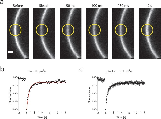
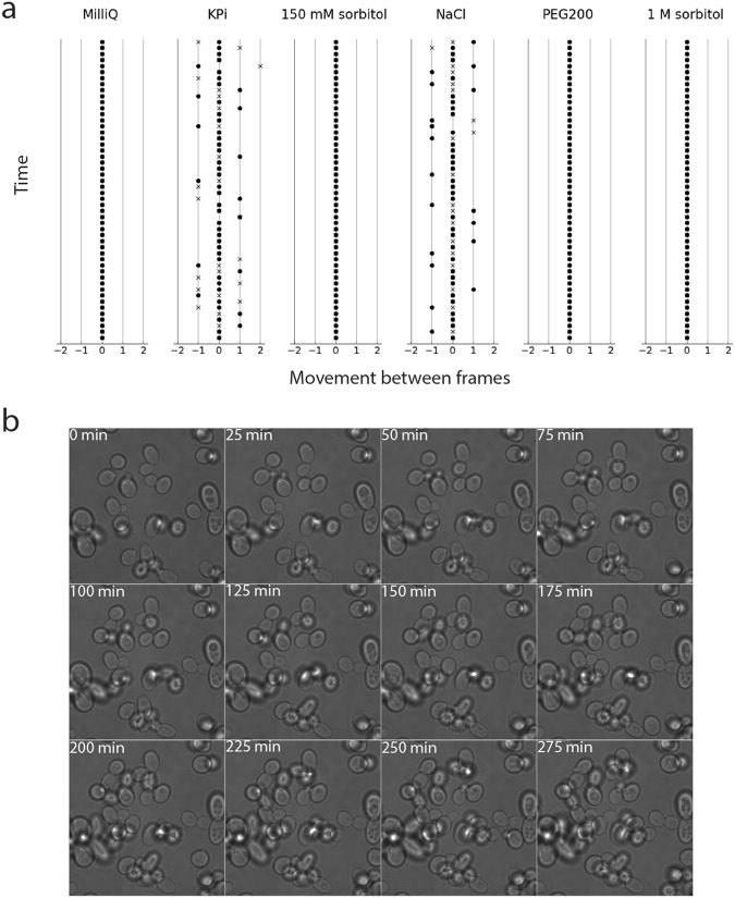
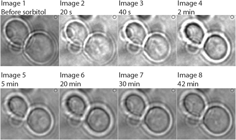
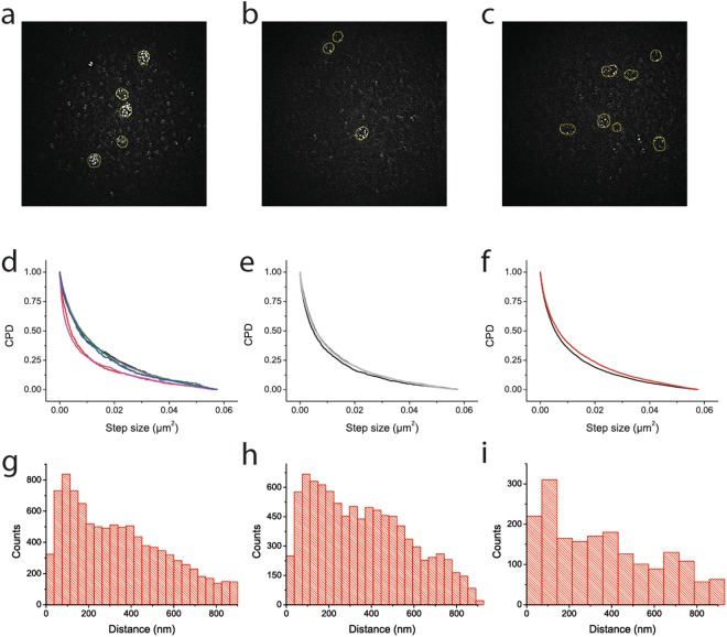
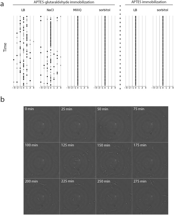
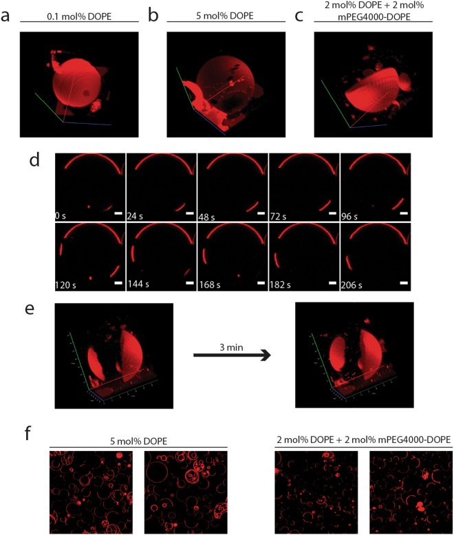
Super-resolution imaging and single-particle tracking require cells to be immobile as any movement reduces the resolution of the measurements. Here, we present a method based on APTES-glutaraldehyde coating of glass surfaces to immobilize cells without compromising their growth. Our method of immobilization is compatible with Saccharomyces cerevisiae, Escherichia coli, and synthetic cells (here, giant-unilamellar vesicles). The method introduces minimal background fluorescence and is suitable for imaging of single particles at high resolution. With S. cerevisiae we benchmarked the method against the commonly used concanavalin A approach. We show by total internal reflection fluorescence microscopy that modifying surfaces with ConA introduces artifacts close to the glass surface, which are not present when immobilizing with the APTES-glutaraldehyde method. We demonstrate validity of the method by measuring the diffusion of membrane proteins in yeast with single-particle tracking and of lipids in giant-unilamellar vesicles with fluorescence recovery after photobleaching. Importantly, the physical properties and shape of the fragile GUVs are not affected upon binding to APTES-glutaraldehyde coated glass. The APTES-glutaraldehyde is a generic method of immobilization that should work with any cell or synthetic system that has primary amines on the surface.
超分辨率成像和单粒子追踪需要细胞处于静止状态,因为任何运动都会降低测量的分辨率。在这里,我们提出了一种基于 APTES-戊二醛对玻璃表面进行涂层处理以固定细胞而不影响其生长的方法。我们的固定方法与酿酒酵母、大肠杆菌和合成细胞(这里是巨大的单层囊泡)兼容。该方法引入的背景荧光最小,适用于高分辨率的单粒子成像。我们使用酿酒酵母对该方法进行了基准测试,与常用的刀豆球蛋白 A 方法进行了比较。我们通过全内反射荧光显微镜表明,用 ConA 修饰表面会在靠近玻璃表面处引入伪影,而在用 APTES-戊二醛方法固定时则不会出现这些伪影。我们通过使用单粒子追踪测量酵母中膜蛋白的扩散以及通过荧光恢复后光漂白测量巨大的单层囊泡中的脂质扩散,证明了该方法的有效性。重要的是,在与 APTES-戊二醛涂层玻璃结合后,脆弱的 GUV 的物理性质和形状不会受到影响。APTES-戊二醛是一种通用的固定化方法,应该适用于任何具有表面伯胺的细胞或合成系统。