• 文献检索
  • 文档翻译
  • 深度研究
  • 学术资讯
  • Suppr Zotero 插件Zotero 插件
  • 邀请有礼
  • 套餐&价格
  • 历史记录
应用&插件
Suppr Zotero 插件Zotero 插件浏览器插件Mac 客户端Windows 客户端微信小程序
定价
高级版会员购买积分包购买API积分包
服务
文献检索文档翻译深度研究API 文档MCP 服务
关于我们
关于 Suppr公司介绍联系我们用户协议隐私条款
关注我们

Suppr 超能文献

核心技术专利:CN118964589B侵权必究
粤ICP备2023148730 号-1Suppr @ 2026

文献检索

告别复杂PubMed语法,用中文像聊天一样搜索,搜遍4000万医学文献。AI智能推荐,让科研检索更轻松。

立即免费搜索

文件翻译

保留排版,准确专业,支持PDF/Word/PPT等文件格式,支持 12+语言互译。

免费翻译文档

深度研究

AI帮你快速写综述,25分钟生成高质量综述,智能提取关键信息,辅助科研写作。

立即免费体验

视紫红质与小型 G 蛋白复合物的晶体结构阐明了 G 蛋白选择性的原理。

Crystal structure of rhodopsin in complex with a mini-G sheds light on the principles of G protein selectivity.

机构信息

Laboratory of Biomolecular Research, Paul Scherrer Institute (PSI), 5232 Villigen PSI, Switzerland.

Department of Biology, ETH Zürich, Wolfgang-Pauli-Strasse 27, 8093 Zürich, Switzerland.

出版信息

Sci Adv. 2018 Sep 19;4(9):eaat7052. doi: 10.1126/sciadv.aat7052. eCollection 2018 Sep.

DOI:10.1126/sciadv.aat7052
PMID:30255144
原文链接:https://pmc.ncbi.nlm.nih.gov/articles/PMC6154990/
Abstract

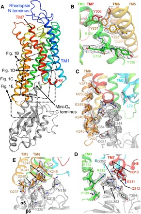
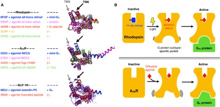
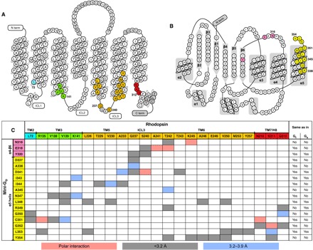
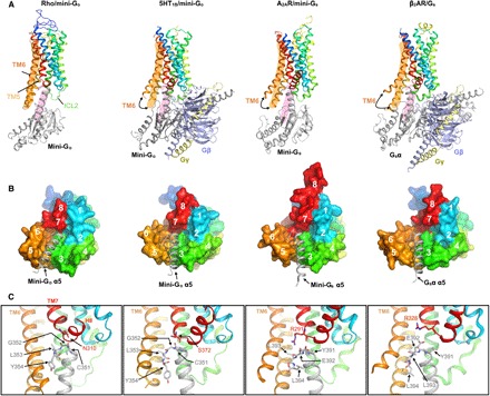
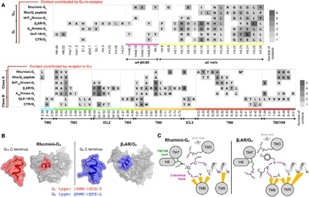
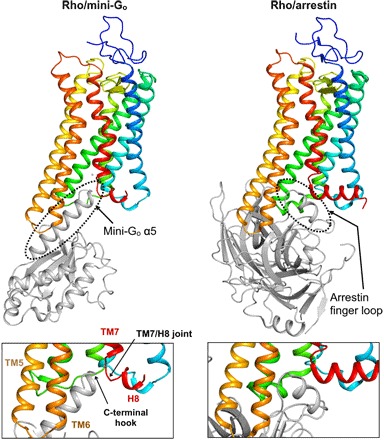
Selective coupling of G protein (heterotrimeric guanine nucleotide-binding protein)-coupled receptors (GPCRs) to specific Gα-protein subtypes is critical to transform extracellular signals, carried by natural ligands and clinical drugs, into cellular responses. At the center of this transduction event lies the formation of a signaling complex between the receptor and G protein. We report the crystal structure of light-sensitive GPCR rhodopsin bound to an engineered mini-G protein. The conformation of the receptor is identical to all previous structures of active rhodopsin, including the complex with arrestin. Thus, rhodopsin seems to adopt predominantly one thermodynamically stable active conformation, effectively acting like a "structural switch," allowing for maximum efficiency in the visual system. Furthermore, our analysis of the well-defined GPCR-G protein interface suggests that the precise position of the carboxyl-terminal "hook-like" element of the G protein (its four last residues) relative to the TM7/helix 8 (H8) joint of the receptor is a significant determinant in selective G protein activation.

摘要

G 蛋白(异三聚体鸟苷酸结合蛋白)偶联受体(GPCR)与特定 Gα-蛋白亚型的选择性偶联对于将天然配体和临床药物携带的细胞外信号转化为细胞反应至关重要。在这种转导事件的中心,受体和 G 蛋白之间形成了一个信号复合物。我们报告了光敏感 GPCR 视紫红质与工程化的迷你 G 蛋白结合的晶体结构。该受体的构象与所有先前的活性视紫红质结构相同,包括与阻滞蛋白的复合物。因此,视紫红质似乎主要采用一种热力学稳定的活性构象,有效地充当“结构开关”,从而在视觉系统中实现最大效率。此外,我们对明确的 GPCR-G 蛋白界面的分析表明,G 蛋白羧基末端“钩状”元件(其最后四个残基)相对于受体 TM7/螺旋 8(H8)连接处的精确位置是选择性 G 蛋白激活的重要决定因素。

https://cdn.ncbi.nlm.nih.gov/pmc/blobs/1037/6154990/78e08448958e/aat7052-F6.jpg
https://cdn.ncbi.nlm.nih.gov/pmc/blobs/1037/6154990/d03316eb604f/aat7052-F1.jpg
https://cdn.ncbi.nlm.nih.gov/pmc/blobs/1037/6154990/840dc3f06923/aat7052-F2.jpg
https://cdn.ncbi.nlm.nih.gov/pmc/blobs/1037/6154990/3d90cbab4ffe/aat7052-F3.jpg
https://cdn.ncbi.nlm.nih.gov/pmc/blobs/1037/6154990/0df0d741b180/aat7052-F4.jpg
https://cdn.ncbi.nlm.nih.gov/pmc/blobs/1037/6154990/e44e4505b203/aat7052-F5.jpg
https://cdn.ncbi.nlm.nih.gov/pmc/blobs/1037/6154990/78e08448958e/aat7052-F6.jpg

相似文献

1
Crystal structure of rhodopsin in complex with a mini-G sheds light on the principles of G protein selectivity.视紫红质与小型 G 蛋白复合物的晶体结构阐明了 G 蛋白选择性的原理。
Sci Adv. 2018 Sep 19;4(9):eaat7052. doi: 10.1126/sciadv.aat7052. eCollection 2018 Sep.
2
Structure and activation of rhodopsin.视紫红质的结构与激活
Acta Pharmacol Sin. 2012 Mar;33(3):291-9. doi: 10.1038/aps.2011.171. Epub 2012 Jan 23.
3
Agonist-induced conformational changes in bovine rhodopsin: insight into activation of G-protein-coupled receptors.激动剂诱导的牛视紫红质构象变化:深入了解G蛋白偶联受体的激活
J Mol Biol. 2008 Oct 3;382(2):539-55. doi: 10.1016/j.jmb.2008.06.084. Epub 2008 Jul 7.
4
Crystal structure of rhodopsin bound to arrestin by femtosecond X-ray laser.通过飞秒X射线激光获得的视紫红质与抑制蛋白结合的晶体结构。
Nature. 2015 Jul 30;523(7562):561-7. doi: 10.1038/nature14656. Epub 2015 Jul 22.
5
Structural features of activated GPCR signaling complexes.激活 G 蛋白偶联受体信号复合物的结构特征。
Curr Opin Struct Biol. 2020 Aug;63:82-89. doi: 10.1016/j.sbi.2020.04.008. Epub 2020 May 30.
6
Conformational selection and equilibrium governs the ability of retinals to bind opsin.构象选择和平衡决定视黄醛与视蛋白结合的能力。
J Biol Chem. 2015 Feb 13;290(7):4304-18. doi: 10.1074/jbc.M114.603134. Epub 2014 Dec 1.
7
Conformational dynamics of helix 8 in the GPCR rhodopsin controls arrestin activation in the desensitization process.GPCR 视紫红质中螺旋 8 的构象动力学控制了脱敏过程中抑制蛋白的激活。
Proc Natl Acad Sci U S A. 2011 Nov 15;108(46):18690-5. doi: 10.1073/pnas.1015461108. Epub 2011 Oct 28.
8
G protein-coupled receptors--recent advances.G蛋白偶联受体——最新进展
Acta Biochim Pol. 2012;59(4):515-29. Epub 2012 Dec 18.
9
A G protein-coupled receptor at work: the rhodopsin model.工作中的 G 蛋白偶联受体:视紫红质模型。
Trends Biochem Sci. 2009 Nov;34(11):540-52. doi: 10.1016/j.tibs.2009.07.005. Epub 2009 Oct 21.
10
Sequence of interactions in receptor-G protein coupling.受体 - G蛋白偶联中的相互作用序列。
J Biol Chem. 2004 Jun 4;279(23):24283-90. doi: 10.1074/jbc.M311166200. Epub 2004 Mar 8.

引用本文的文献

1
Deprotonation of retinal Schiff base and structural dynamics in the early photoreaction of primate blue cone visual pigment.灵长类动物蓝锥视觉色素早期光反应中视黄醛席夫碱的去质子化及结构动力学
Biophys J. 2025 Jun 17;124(12):2070-2081. doi: 10.1016/j.bpj.2025.05.004. Epub 2025 May 7.
2
Active state structures of a bistable visual opsin bound to G proteins.与 G 蛋白结合的双稳态视觉视蛋白的活性状态结构。
Nat Commun. 2024 Oct 16;15(1):8928. doi: 10.1038/s41467-024-53208-2.
3
Molecular mechanism of distinct chemokine engagement and functional divergence of the human Duffy antigen receptor.

本文引用的文献

1
Cryo-EM structure of the serotonin 5-HT receptor coupled to heterotrimeric G.血清素 5-HT 受体与异三聚体 G 偶联的冷冻电镜结构。
Nature. 2018 Jun;558(7711):620-623. doi: 10.1038/s41586-018-0241-9. Epub 2018 Jun 20.
2
Ligand channel in pharmacologically stabilized rhodopsin.药理学稳定视紫红质中的配体通道。
Proc Natl Acad Sci U S A. 2018 Apr 3;115(14):3640-3645. doi: 10.1073/pnas.1718084115. Epub 2018 Mar 19.
3
Mini G protein probes for active G protein-coupled receptors (GPCRs) in live cells.用于活细胞中活性 G 蛋白偶联受体 (GPCR) 的微型 G 蛋白探针。
人 Duffy 抗原受体结合不同趋化因子的分子机制及其功能分化。
Cell. 2024 Aug 22;187(17):4751-4769.e25. doi: 10.1016/j.cell.2024.07.005. Epub 2024 Jul 31.
4
Ligand Binding Affinity Prediction for Membrane Proteins with Alchemical Free Energy Calculation Methods.基于热力学积分方法的膜蛋白配体结合亲和力预测。
J Chem Inf Model. 2024 Jul 22;64(14):5671-5679. doi: 10.1021/acs.jcim.4c00764. Epub 2024 Jul 3.
5
Exploring GPCR conformational dynamics using single-molecule fluorescence.利用单分子荧光技术探索 G 蛋白偶联受体构象动力学。
Methods. 2024 Jun;226:35-48. doi: 10.1016/j.ymeth.2024.03.011. Epub 2024 Apr 10.
6
A pedigree with retinitis pigmentosa and its concomitant ophthalmic diseases.一个患有视网膜色素变性及其伴发眼科疾病的家系。
Int J Ophthalmol. 2023 Dec 18;16(12):1962-1970. doi: 10.18240/ijo.2023.12.07. eCollection 2023.
7
A rapid, tag-free way to purify functional GPCRs.一种快速、无标签的方法来纯化功能性 G 蛋白偶联受体。
J Biol Chem. 2024 Jan;300(1):105558. doi: 10.1016/j.jbc.2023.105558. Epub 2023 Dec 12.
8
Gαs slow conformational transition upon GTP binding and a novel Gαs regulator.GTP结合时Gαs的缓慢构象转变及一种新型Gαs调节剂。
iScience. 2023 Apr 8;26(5):106603. doi: 10.1016/j.isci.2023.106603. eCollection 2023 May 19.
9
A visual opsin from jellyfish enables precise temporal control of G protein signalling.水母的一种视蛋白能够精确控制 G 蛋白信号转导。
Nat Commun. 2023 Apr 28;14(1):2450. doi: 10.1038/s41467-023-38231-z.
10
Functional optimization of light-activatable Opto-GPCRs: Illuminating the importance of the proximal C-terminus in G-protein specificity.光激活型光遗传学G蛋白偶联受体(Opto-GPCRs)的功能优化:揭示近侧C末端在G蛋白特异性中的重要性
Front Cell Dev Biol. 2023 Mar 1;11:1053022. doi: 10.3389/fcell.2023.1053022. eCollection 2023.
J Biol Chem. 2018 May 11;293(19):7466-7473. doi: 10.1074/jbc.RA118.001975. Epub 2018 Mar 9.
4
G- and G-coupled GPCRs show different modes of G-protein binding.G 蛋白偶联受体(GPCRs)中的 G 蛋白偶联和 G 蛋白偶联型受体(GPCRs)表现出不同的 G 蛋白结合模式。
Proc Natl Acad Sci U S A. 2018 Mar 6;115(10):2383-2388. doi: 10.1073/pnas.1721896115. Epub 2018 Feb 20.
5
Single-molecule analysis of ligand efficacy in βAR-G-protein activation.β肾上腺素能受体(βAR)-G蛋白激活中配体效能的单分子分析
Nature. 2017 Jul 6;547(7661):68-73. doi: 10.1038/nature22354. Epub 2017 Jun 7.
6
Selectivity determinants of GPCR-G-protein binding.G蛋白偶联受体(GPCR)与G蛋白结合的选择性决定因素。
Nature. 2017 May 18;545(7654):317-322. doi: 10.1038/nature22070. Epub 2017 May 10.
7
Expression and Purification of Mini G Proteins from .来自……的小G蛋白的表达与纯化
Bio Protoc. 2017 Apr 20;7(8). doi: 10.21769/BioProtoc.2235.
8
Mini-G proteins: Novel tools for studying GPCRs in their active conformation.小G蛋白:研究处于活性构象的G蛋白偶联受体的新型工具。
PLoS One. 2017 Apr 20;12(4):e0175642. doi: 10.1371/journal.pone.0175642. eCollection 2017.
9
Conformational equilibria of light-activated rhodopsin in nanodiscs.纳米盘状结构中光激活视紫红质的构象平衡。
Proc Natl Acad Sci U S A. 2017 Apr 18;114(16):E3268-E3275. doi: 10.1073/pnas.1620405114. Epub 2017 Apr 3.
10
Polder maps: improving OMIT maps by excluding bulk solvent.Polder 图:通过排除主体溶剂来改进 OMIT 图。
Acta Crystallogr D Struct Biol. 2017 Feb 1;73(Pt 2):148-157. doi: 10.1107/S2059798316018210.