Department of Infection Biology, Institute of Infection and Global Health and School of Veterinary Science, Faculty of Health and Life Sciences, University of Liverpool, Merseyside, UK.
Institute of Integrative Biology, University of Liverpool, Liverpool, UK.
Cell Tissue Res. 2019 Feb;375(2):409-424. doi: 10.1007/s00441-018-2924-9. Epub 2018 Sep 26.
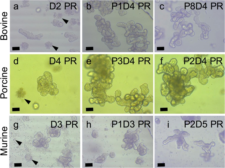
The in vitro 3D culture of intestinal epithelium is a valuable resource in the study of its function. Organoid culture exploits stem cells' ability to regenerate and produce differentiated epithelium. Intestinal organoid models from rodent or human tissue are widely available whereas large animal models are not. Livestock enteric and zoonotic diseases elicit significant morbidity and mortality in animal and human populations. Therefore, livestock species-specific models may offer novel insights into host-pathogen interactions and disease responses. Bovine and porcine jejunum were obtained from an abattoir and their intestinal crypts isolated, suspended in Matrigel, cultured, cryopreserved and resuscitated. 'Rounding' of crypts occurred followed by budding and then enlargement of the organoids. Epithelial cells were characterised using immunofluorescent staining and confocal microscopy. Organoids were successfully infected with Toxoplasma gondii or Salmonella typhimurium. This 3D organoid model offers a long-term, renewable resource for investigating species-specific intestinal infections with a variety of pathogens.
肠上皮的体外 3D 培养是研究其功能的宝贵资源。类器官培养利用了干细胞的再生和产生分化上皮的能力。来自啮齿动物或人类组织的肠类器官模型广泛可用,而大型动物模型则不可用。牲畜肠道和人畜共患病在动物和人群中引起了很高的发病率和死亡率。因此,特定于牲畜的模型可能为宿主-病原体相互作用和疾病反应提供新的见解。从屠宰场获得牛和猪的空肠,并分离其肠隐窝,悬浮在 Matrigel 中,培养、冷冻保存和复苏。隐窝“变圆”,然后出芽,然后类器官增大。使用免疫荧光染色和共聚焦显微镜对上皮细胞进行了特征描述。类器官成功感染了刚地弓形虫或鼠伤寒沙门氏菌。这种 3D 类器官模型为研究具有各种病原体的特定于物种的肠道感染提供了长期的、可再生的资源。