Department of Orthopedics, The Affiliated Huaian No. 1 People's Hospital of Nanjing Medical University, Huaian, Jiangsu, China (mainland).
Department of Orthopedics, Nanjing First Hospital, Nanjing Medical University, Nanjing, Jiangsu, China (mainland).
Med Sci Monit. 2018 Sep 30;24:6934-6945. doi: 10.12659/MSM.911770.
BACKGROUND The aim of this study was to design and test a novel composite scaffold with antibacterial efficacy for treating bone infections using a three-dimensional (3D) printed poly(ε-caprolactone) (PCL) scaffold coated with polydopamine (PDA) for the adsorption of polylactic acid-glycolic acid (PLGA) microspheres loaded with vancomycin. MATERIAL AND METHODS Vancomycin-loaded PLGA microspheres were produced by the double-emulsion method, and microsphere morphology, drug-loading dosage, encapsulation efficiency, average diameter, and release characteristics were examined. Composite scaffolds were prepared by adsorption of the microspheres on PDA-coated, 3D-printed PCL scaffolds, and scaffold morphology, biocompatibility, vancomycin release, and antibacterial efficacy were evaluated. RESULTS The vancomycin-loaded microspheres were smooth, round, uniform in size, and had no adhesion phenomenon, and exhibited sustained release of vancomycin from the microspheres for more than 4 weeks. Upon modification with PDA, the PCL scaffold changed from white to black, and after microsphere adsorption, dot-like white particles were seen. On scanning electron microscopy, PDA particles were observed on the PCL/PDA composite scaffolds, and PLGA microspheres were evenly dispersed over the PDA coating on the PCL/PDA/PLGA composite scaffolds. Cell viability assays showed that the adhesion and proliferation of rabbit bone mesenchymal stem cells were greater on the PCL/PDA scaffolds than on unmodified PCL scaffolds. Microsphere adsorption had no significant effect on cell proliferation. In vitro release of vancomycin from the composite scaffolds was observed for more than 4 weeks, and observation of the inhibition zone on agar plates of Staphylococcus aureus showed that the scaffolds maintained their antibacterial effect for more than 4 weeks. CONCLUSIONS The 3D-printed, PDA-coated PCL scaffold carrying vancomycin-loaded PLGA microspheres exhibited good biocompatibility and a sustained antibacterial effect in vitro.
本研究旨在设计并测试一种新型复合支架,该支架具有抗菌功效,可用于治疗骨感染。采用 3D 打印聚己内酯(PCL)支架,其表面涂有多巴胺(PDA),以吸附载万古霉素的聚乳酸-羟基乙酸(PLGA)微球。
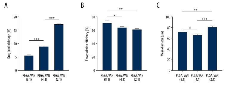
采用复乳法制备载万古霉素 PLGA 微球,考察微球的形态、载药量、包封率、平均粒径和释放特性。通过吸附载万古霉素 PLGA 微球制备复合支架,考察支架的形态、生物相容性、万古霉素释放和抗菌效果。
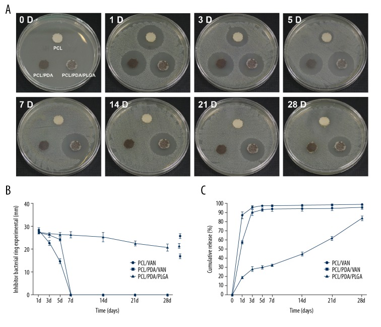
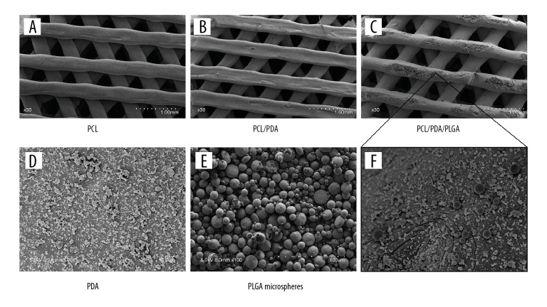
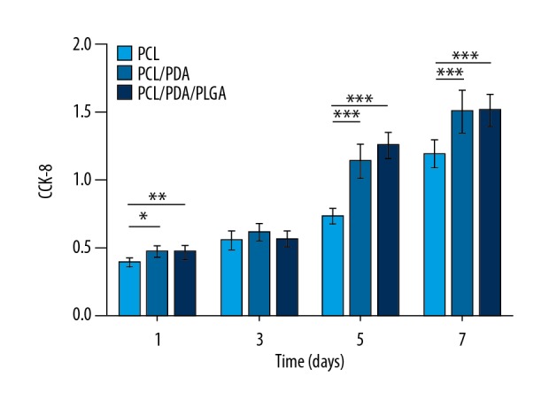
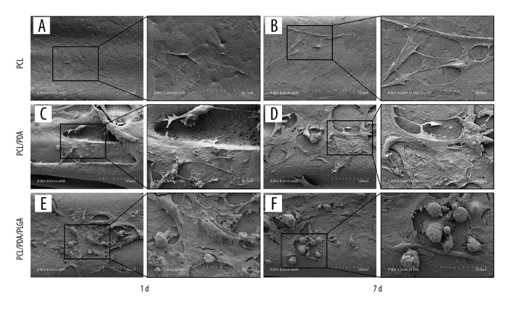
载万古霉素微球呈光滑、圆整、粒径均一、无粘连现象,微球载药后能够持续释放万古霉素 4 周以上。PCL 支架经 PDA 修饰后由白色变为黑色,吸附微球后可见点状白色颗粒。扫描电镜观察到 PCL/PDA 复合支架上有 PDA 颗粒,PCL/PDA/PLGA 复合支架上的 PDA 涂层均匀分布着 PLGA 微球。细胞活力实验表明,兔骨髓间充质干细胞在 PCL/PDA 支架上的黏附和增殖能力强于未改性 PCL 支架。微球吸附对细胞增殖无明显影响。复合支架中万古霉素体外释放超过 4 周,琼脂平板抑菌圈观察到支架对金黄色葡萄球菌的抗菌作用超过 4 周。
载万古霉素 PLGA 微球的 3D 打印 PDA 涂层 PCL 支架具有良好的生物相容性和体外持续抗菌效果。