• 文献检索
  • 文档翻译
  • 深度研究
  • 学术资讯
  • Suppr Zotero 插件Zotero 插件
  • 邀请有礼
  • 套餐&价格
  • 历史记录
应用&插件
Suppr Zotero 插件Zotero 插件浏览器插件Mac 客户端Windows 客户端微信小程序
定价
高级版会员购买积分包购买API积分包
服务
文献检索文档翻译深度研究API 文档MCP 服务
关于我们
关于 Suppr公司介绍联系我们用户协议隐私条款
关注我们

Suppr 超能文献

核心技术专利:CN118964589B侵权必究
粤ICP备2023148730 号-1Suppr @ 2026

文献检索

告别复杂PubMed语法,用中文像聊天一样搜索,搜遍4000万医学文献。AI智能推荐,让科研检索更轻松。

立即免费搜索

文件翻译

保留排版,准确专业,支持PDF/Word/PPT等文件格式,支持 12+语言互译。

免费翻译文档

深度研究

AI帮你快速写综述,25分钟生成高质量综述,智能提取关键信息,辅助科研写作。

立即免费体验

使用基于色相-饱和度-亮度的表面创建来简化体积多通道图像细胞术

Streamlining volumetric multi-channel image cytometry using hue-saturation-brightness-based surface creation.

作者信息

Tan Yingrou, Li Jackson Liang Yao, Goh Chi Ching, Lee Bernett Teck Kwong, Kwok Immanuel Weng Han, Ng Wei Jie, Evrard Maximilien, Poidinger Michael, Tey Hong Liang, Ng Lai Guan

机构信息

Singapore Immunology Network (SIgN), A*STAR (Agency for Science, Technology and Research), Biopolis, 138648, Singapore, Singapore.

National Skin Centre, 1 Mandalay Road, 308205, Singapore, Singapore.

出版信息

Commun Biol. 2018 Sep 6;1:136. doi: 10.1038/s42003-018-0139-y. eCollection 2018.

DOI:10.1038/s42003-018-0139-y
PMID:30272015
原文链接:https://pmc.ncbi.nlm.nih.gov/articles/PMC6127105/
Abstract

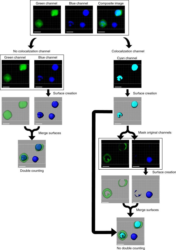
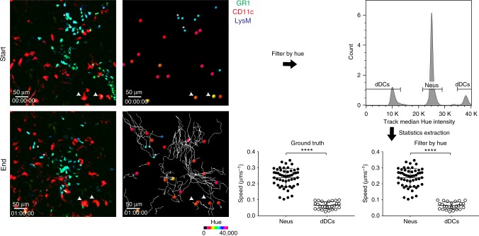
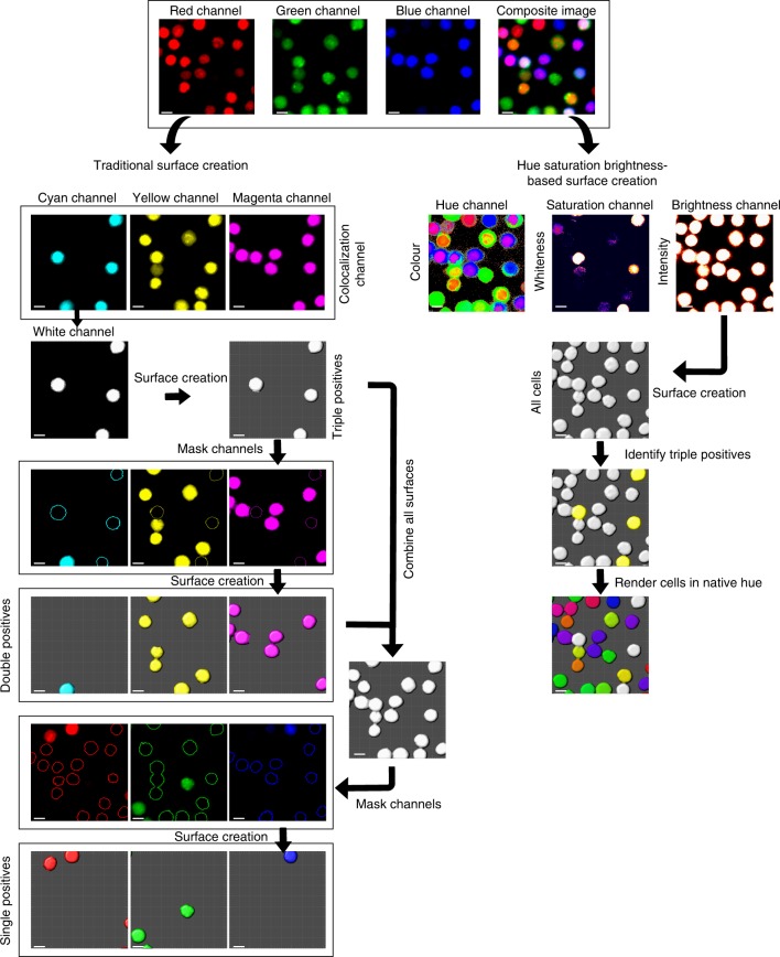
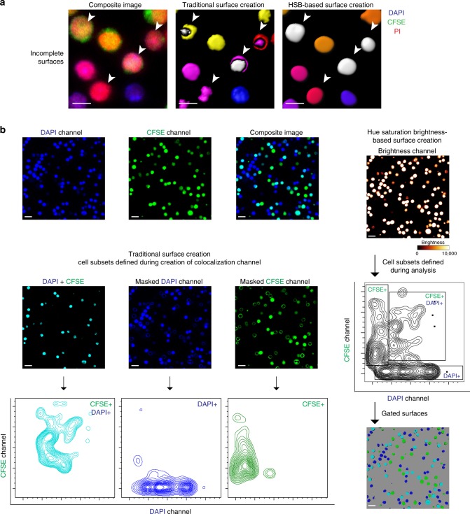
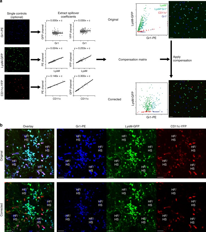
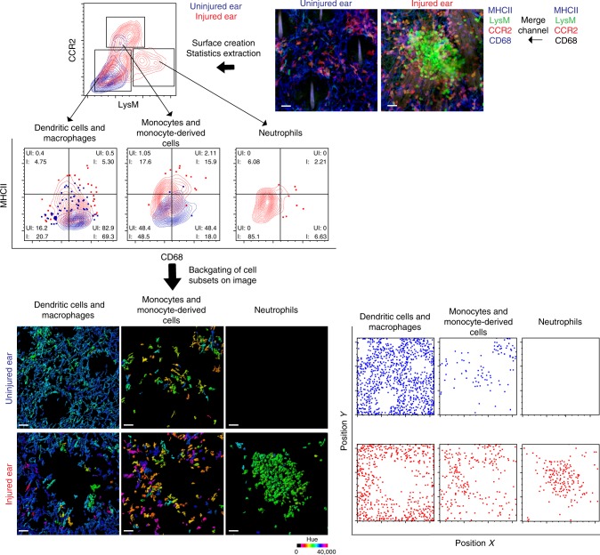
Image cytometry is the process of converting image data to flow cytometry-style plots, and it usually requires computer-aided surface creation to extract out statistics for cells or structures. One way of dealing with structures stained with multiple markers in three-dimensional images, is carrying out multiple rounds of channel co-localization and image masking before surface creation, which is cumbersome and laborious. We propose the application of the hue-saturation-brightness color space to streamline this process, which produces complete surfaces, and allows the user to have a global view of the data before flexibly defining cell subsets. Spectral compensation can also be performed after surface creation to accurately resolve different signals. We demonstrate the utility of this workflow in static and dynamic imaging datasets of a needlestick injury on the mouse ear, and we believe this scalable and intuitive approach will improve the ease of performing histocytometry on biological samples.

摘要

图像细胞术是将图像数据转换为流式细胞术风格图表的过程,通常需要借助计算机辅助创建表面来提取细胞或结构的统计数据。处理三维图像中用多种标记物染色的结构的一种方法是,在创建表面之前进行多轮通道共定位和图像掩膜,这既繁琐又费力。我们建议应用色相-饱和度-亮度颜色空间来简化这一过程,该方法能生成完整的表面,并允许用户在灵活定义细胞亚群之前对数据有一个全局的了解。在创建表面之后还可以进行光谱补偿,以准确解析不同的信号。我们在小鼠耳部针刺损伤的静态和动态成像数据集中展示了此工作流程的实用性,并且我们相信这种可扩展且直观的方法将提高对生物样品进行组织细胞计数的简便性。

https://cdn.ncbi.nlm.nih.gov/pmc/blobs/97d5/6127105/d6e41cbb0dd7/42003_2018_139_Fig6_HTML.jpg
https://cdn.ncbi.nlm.nih.gov/pmc/blobs/97d5/6127105/46d908a3cb14/42003_2018_139_Fig1_HTML.jpg
https://cdn.ncbi.nlm.nih.gov/pmc/blobs/97d5/6127105/28f3481e6e45/42003_2018_139_Fig2_HTML.jpg
https://cdn.ncbi.nlm.nih.gov/pmc/blobs/97d5/6127105/339870278bbe/42003_2018_139_Fig3_HTML.jpg
https://cdn.ncbi.nlm.nih.gov/pmc/blobs/97d5/6127105/b7e38d3580e7/42003_2018_139_Fig4_HTML.jpg
https://cdn.ncbi.nlm.nih.gov/pmc/blobs/97d5/6127105/989c3e719b5e/42003_2018_139_Fig5_HTML.jpg
https://cdn.ncbi.nlm.nih.gov/pmc/blobs/97d5/6127105/d6e41cbb0dd7/42003_2018_139_Fig6_HTML.jpg

相似文献

1
Streamlining volumetric multi-channel image cytometry using hue-saturation-brightness-based surface creation.使用基于色相-饱和度-亮度的表面创建来简化体积多通道图像细胞术
Commun Biol. 2018 Sep 6;1:136. doi: 10.1038/s42003-018-0139-y. eCollection 2018.
2
Visualization of three-way comparisons of omics data.组学数据的三方比较可视化。
BMC Bioinformatics. 2007 Mar 5;8:72. doi: 10.1186/1471-2105-8-72.
3
An Extension of Reversible Image Enhancement Processing for Saturation and Brightness Contrast.用于饱和度和亮度对比度的可逆图像增强处理扩展
J Imaging. 2022 Jan 28;8(2):27. doi: 10.3390/jimaging8020027.
4
Transformation of a high-dimensional color space for material classification.用于材料分类的高维颜色空间变换。
J Opt Soc Am A Opt Image Sci Vis. 2017 Apr 1;34(4):523-532. doi: 10.1364/JOSAA.34.000523.
5
[Segmentation and cavity filling of color image from stained trabecular sections based on HSV space].基于HSV空间的染色小梁切片彩色图像分割与空洞填充
Sheng Wu Yi Xue Gong Cheng Xue Za Zhi. 2012 Apr;29(2):260-3.
6
Radiometric Compensation of Images Projected on Non-White Surfaces by Exploiting Chromatic Adaptation and Perceptual Anchoring.利用颜色适应和感知锚定对非白色表面投影图像进行辐射补偿。
IEEE Trans Image Process. 2017 Jan;26(1):147-159. doi: 10.1109/TIP.2016.2592799. Epub 2016 Jul 18.
7
Extracting dominant textures in real time with multi-scale hue-saturation-intensity histograms.实时提取多尺度色调-饱和度-强度直方图中的优势纹理。
IEEE Trans Image Process. 2013 Nov;22(11):4237-48. doi: 10.1109/TIP.2013.2271426. Epub 2013 Jun 27.
8
An open-source solution for advanced imaging flow cytometry data analysis using machine learning.一种使用机器学习进行高级成像流式细胞术数据分析的开源解决方案。
Methods. 2017 Jan 1;112:201-210. doi: 10.1016/j.ymeth.2016.08.018. Epub 2016 Sep 2.
9
Color and emotion: effects of hue, saturation, and brightness.颜色与情感:色调、饱和度和亮度的影响
Psychol Res. 2018 Sep;82(5):896-914. doi: 10.1007/s00426-017-0880-8. Epub 2017 Jun 13.
10
Frequency difference gating: a multivariate method for identifying subsets that differ between samples.频率差异门控:一种用于识别样本间存在差异的子集的多变量方法。
Cytometry. 2001 Sep 1;45(1):56-64. doi: 10.1002/1097-0320(20010901)45:1<56::aid-cyto1144>3.0.co;2-9.

引用本文的文献

1
Divergent Forms of Pyroplastic: Lessons Learned from the M/V Ship Fire.热塑性塑料的不同形式:从“M/V 船舶火灾”中学到的经验教训
ACS Environ Au. 2022 Jul 29;2(5):467-479. doi: 10.1021/acsenvironau.2c00020. eCollection 2022 Sep 21.
2
Pancreatic β cell-selective zinc transporter 8 insufficiency accelerates diabetes associated with islet amyloidosis.胰岛β细胞选择性锌转运体 8 不足加速与胰岛淀粉样变相关的糖尿病。
JCI Insight. 2021 May 24;6(10):143037. doi: 10.1172/jci.insight.143037.
3
Spatiotemporal regulation of type I interferon expression determines the antiviral polarization of CD4 T cells.

本文引用的文献

1
Multiplex, quantitative cellular analysis in large tissue volumes with clearing-enhanced 3D microscopy (C3D).利用增强清除的 3D 显微镜(C3D)进行多重、定量细胞分析在大组织体积中。
Proc Natl Acad Sci U S A. 2017 Aug 29;114(35):E7321-E7330. doi: 10.1073/pnas.1708981114. Epub 2017 Aug 14.
2
MIST: Accurate and Scalable Microscopy Image Stitching Tool with Stage Modeling and Error Minimization.MIST:具有阶段建模和误差最小化的准确且可扩展的显微镜图像拼接工具。
Sci Rep. 2017 Jul 10;7(1):4988. doi: 10.1038/s41598-017-04567-y.
3
Microbiota-Dependent Sequelae of Acute Infection Compromise Tissue-Specific Immunity.
I 型干扰素表达的时空调节决定了 CD4 T 细胞的抗病毒极化。
Nat Immunol. 2020 Mar;21(3):321-330. doi: 10.1038/s41590-020-0596-6. Epub 2020 Feb 17.
4
Capturing the Fantastic Voyage of Monocytes Through Time and Space.捕捉单核细胞穿越时空的奇幻之旅。
Front Immunol. 2019 Apr 16;10:834. doi: 10.3389/fimmu.2019.00834. eCollection 2019.
急性感染的微生物群依赖性后遗症损害组织特异性免疫。
Cell. 2015 Oct 8;163(2):354-66. doi: 10.1016/j.cell.2015.08.030.
4
Strategically localized dendritic cells promote rapid T cell responses to lymph-borne particulate antigens.策略性定位树突状细胞促进淋巴源性颗粒抗原引发的快速 T 细胞应答。
Immunity. 2015 Jan 20;42(1):172-85. doi: 10.1016/j.immuni.2014.12.024. Epub 2014 Dec 27.
5
Multiplexed immunohistochemistry, imaging, and quantitation: a review, with an assessment of Tyramide signal amplification, multispectral imaging and multiplex analysis.多重免疫组织化学、成像与定量分析:综述,兼对酪胺信号放大、多光谱成像及多重分析的评估
Methods. 2014 Nov;70(1):46-58. doi: 10.1016/j.ymeth.2014.08.016. Epub 2014 Sep 19.
6
Histo-cytometry: a method for highly multiplex quantitative tissue imaging analysis applied to dendritic cell subset microanatomy in lymph nodes.组织细胞计量术:一种高多重定量组织成像分析方法,应用于淋巴结树突状细胞亚群的微观解剖结构。
Immunity. 2012 Aug 24;37(2):364-76. doi: 10.1016/j.immuni.2012.07.011. Epub 2012 Aug 2.
7
Fiji: an open-source platform for biological-image analysis.斐济:一个用于生物影像分析的开源平台。
Nat Methods. 2012 Jun 28;9(7):676-82. doi: 10.1038/nmeth.2019.
8
Visualization of image data from cells to organisms.从细胞到生物体的图像数据可视化。
Nat Methods. 2010 Mar;7(3 Suppl):S26-41. doi: 10.1038/nmeth.1431.
9
CellProfiler: image analysis software for identifying and quantifying cell phenotypes.CellProfiler:用于识别和量化细胞表型的图像分析软件。
Genome Biol. 2006;7(10):R100. doi: 10.1186/gb-2006-7-10-r100. Epub 2006 Oct 31.
10
Microscopy-based multicolor tissue cytometry at the single-cell level.基于显微镜的单细胞水平多色组织细胞计数法。
Cytometry A. 2004 Jun;59(2):182-90. doi: 10.1002/cyto.a.20052.