Department of Zoology, College of Science, King Saud University, Riyadh, Saudi Arabia,
Int J Nanomedicine. 2018 Sep 24;13:5685-5699. doi: 10.2147/IJN.S165448. eCollection 2018.
Graphene oxide nanoparticles have been widely used in industry and biomedical fields due to their unique physicochemical properties. However, comparative cytotoxicity of silver-doped reduced graphene oxide (rGO-Ag) nanoparticles on normal and cancerous liver cells has not been well studied yet.
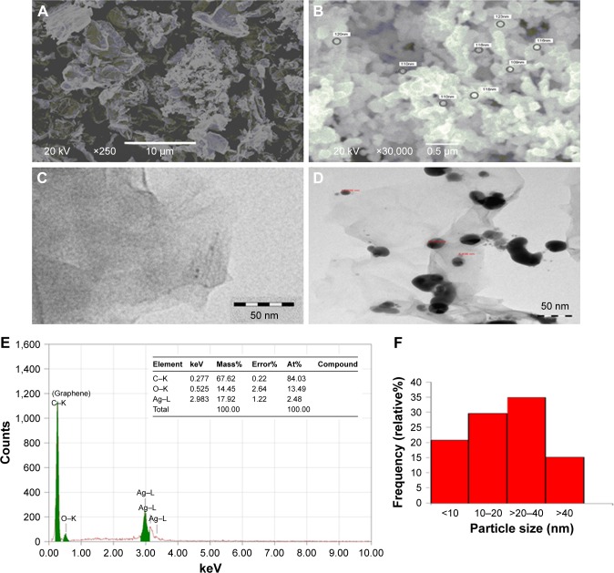
This study aimed at determining the toxic potential of rGO-Ag nanocomposite on human liver normal (CHANG) and cancer (HepG2) cells. The rGO-Ag nanocomposite was characterized by using different advanced instruments, namely, dynamic light scattering, scanning electron microscope, and transmission electron microscope.
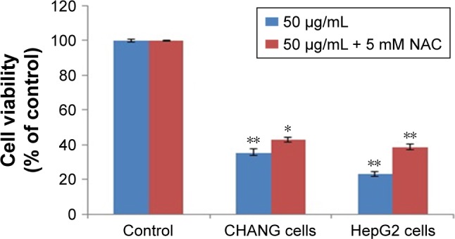
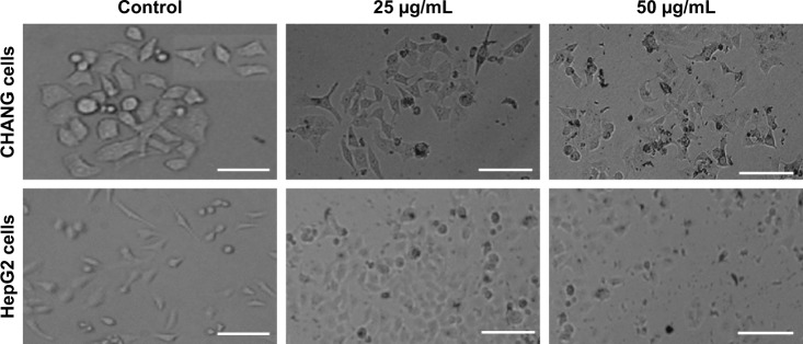
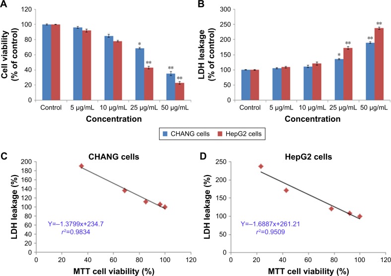
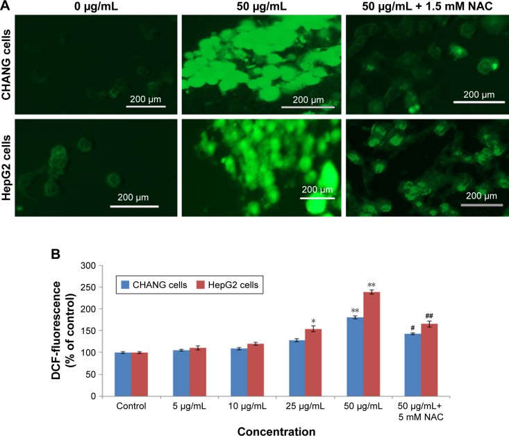
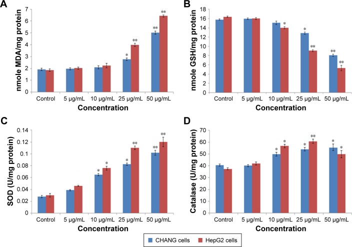
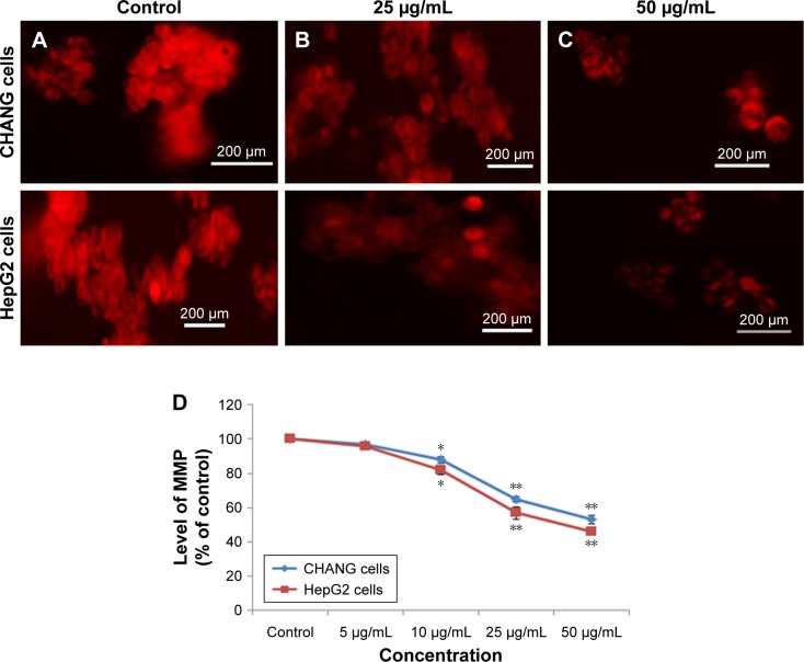
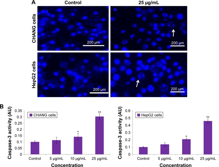
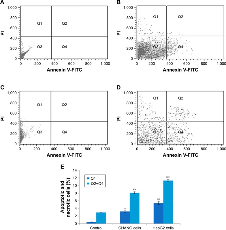
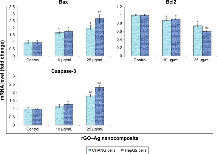
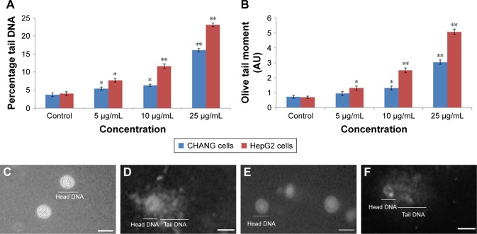
The rGO-Ag nanocomposite reduced cell viability and impaired cell membrane integrity of CHANG and HepG2 cells in a dose-dependent manner. Additionally, it induced reactive oxygen species generation and reduced mitochondrial membrane potential in both cells in a dose-dependent manner. Moreover, the activity of oxidative enzymes such as lipid peroxide, superoxide dismutase, and catalase were increased and glutathione was reduced in both cells exposed to rGO-Ag nanocomposite. Pretreatment with -acetylcysteine inhibited cytotoxicity and reactive oxygen species generation in CHANG and HepG2 cells exposed to rGO-Ag nanocomposite (50 µg/mL). DNA damage was determined by Comet assay and maximum DNA damage occurred at rGO-Ag nanocomposite (25 µg/mL) for 24 h. It is also valuable to inform that HepG2 cells appear to be slightly more susceptible to rGO-Ag nanocomposite exposure than CHANG cells.
This result provides a basic comparative toxic effect of rGO-Ag nanocomposite on hepatic normal and cancerous liver cells.
由于具有独特的物理化学性质,氧化石墨烯纳米粒子已被广泛应用于工业和生物医学领域。然而,银掺杂还原氧化石墨烯(rGO-Ag)纳米粒子对正常和肝癌细胞的比较细胞毒性尚未得到很好的研究。
本研究旨在确定 rGO-Ag 纳米复合材料对人正常肝(CHANG)和肝癌(HepG2)细胞的潜在毒性。使用不同的先进仪器,即动态光散射、扫描电子显微镜和透射电子显微镜对 rGO-Ag 纳米复合材料进行了表征。
rGO-Ag 纳米复合材料以剂量依赖的方式降低了 CHANG 和 HepG2 细胞的细胞活力并破坏了细胞膜的完整性。此外,它还以剂量依赖的方式诱导了两种细胞中活性氧的产生和线粒体膜电位的降低。此外,暴露于 rGO-Ag 纳米复合材料的两种细胞中的氧化酶活性(如脂质过氧化物、超氧化物歧化酶和过氧化氢酶)增加,谷胱甘肽减少。在用 -乙酰半胱氨酸预处理后,暴露于 rGO-Ag 纳米复合材料(50μg/mL)的 CHANG 和 HepG2 细胞的细胞毒性和活性氧的产生被抑制。彗星试验测定 DNA 损伤,在 rGO-Ag 纳米复合材料(25μg/mL)暴露 24 小时时发生最大 DNA 损伤。值得注意的是,与 CHANG 细胞相比,HepG2 细胞似乎对 rGO-Ag 纳米复合材料的暴露更为敏感。
该结果提供了 rGO-Ag 纳米复合材料对正常和肝癌细胞的基本比较毒性作用。