Protein Transport and Secretion Unit, IRCCS Ospedale San Raffaele/Università Vita-Salute San Raffaele, 20132, Milan, Italy.
Cell Biology Unit, Ospedale Policlinico San Martino, 16132, Genoa, Italy.
Cell Death Dis. 2018 Oct 23;9(11):1088. doi: 10.1038/s41419-018-1121-9.
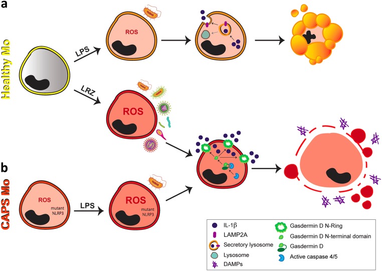
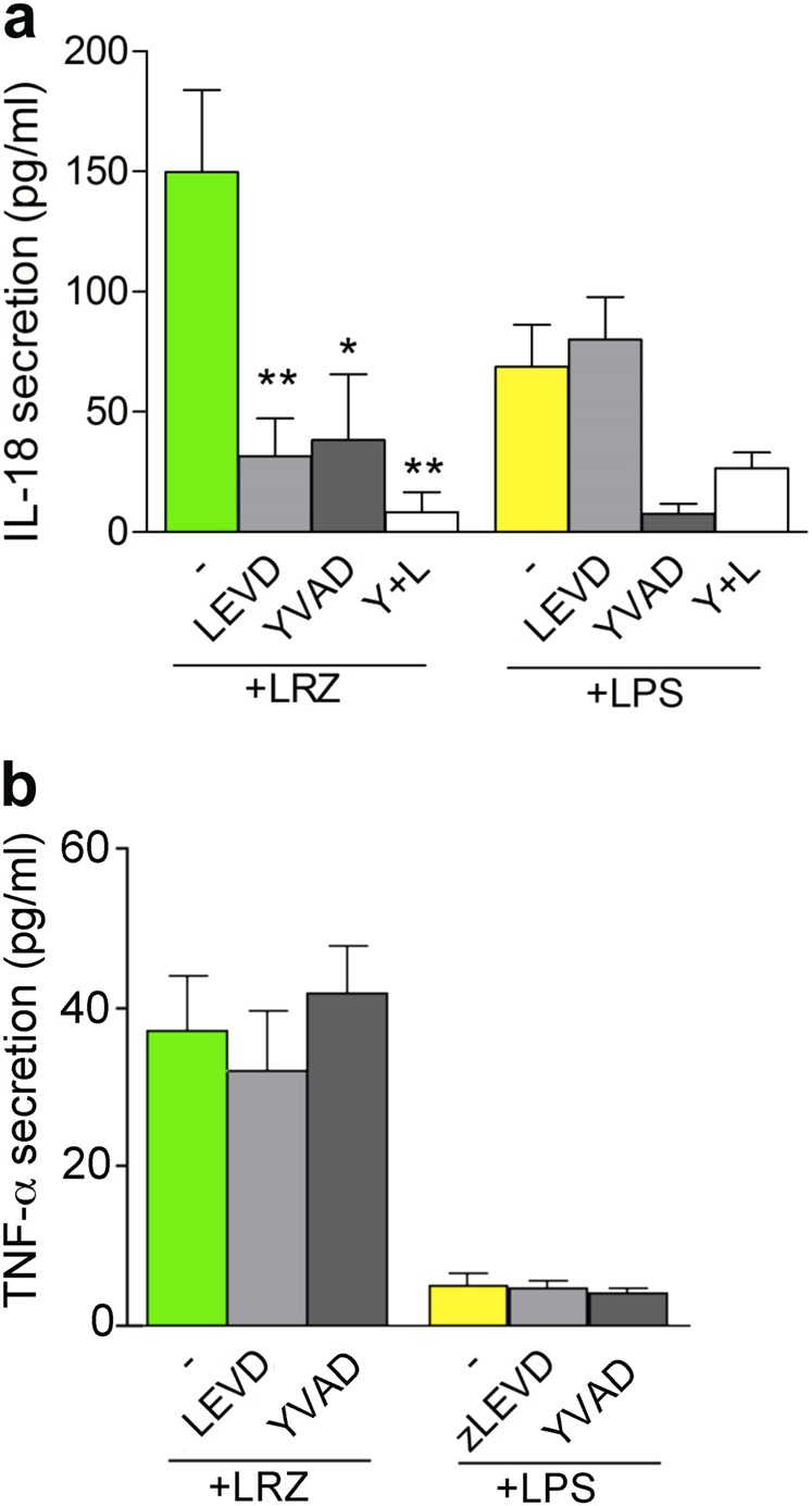
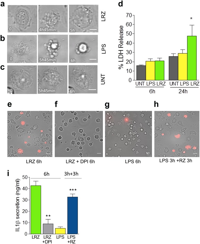
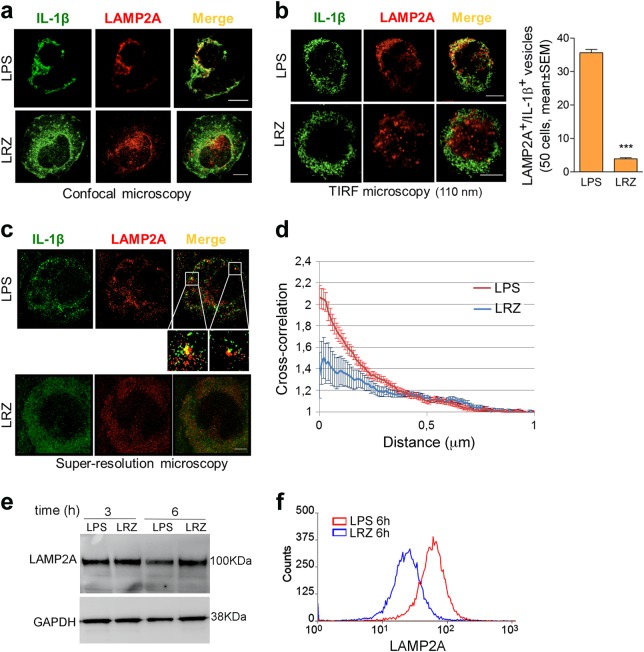
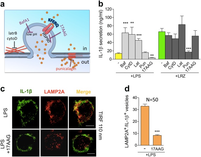
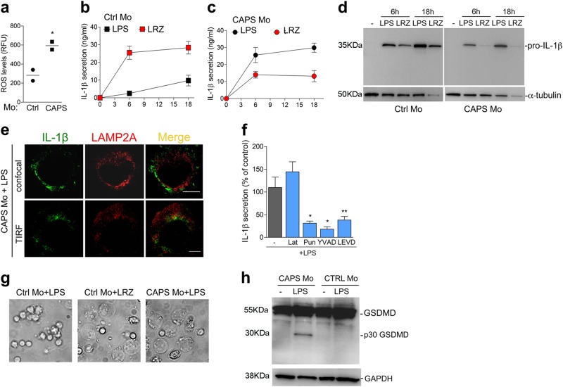
IL-1β is an essential cytokine, but its release needs to be strictly controlled to avoid severe inflammatory manifestations. Lacking a signal sequence, IL-1β does not follow the endoplasmic reticulum-Golgi route. Several pathways have been proposed to mediate its release. One involves the translocation of pro-IL-1β into intracellular vesicles of lysosomal origin that eventually fuse with the plasma membrane. Another exploits pores formed on the plasma membrane upon proteolytic cleavage of gasdermin D (GSDMD). Here we investigated how primary monocytes-the main source of IL-1β in humans-control IL-1β release in response to pro-inflammatory stimuli of increasing intensity and found that two different routes are induced depending on the strength of activation. Triggering of Toll-like receptor 4 (TLR4) by LPS induces slow IL-1β release through LAMP2A vesicles. In contrast, the simultaneous stimulation of TLR2, TLR4 and TLR7/8 drives high levels of ROS, GSDMD cleavage and faster IL-1β secretion. Drugs blocking ROS production prevent GSDMD cleavage supporting a role of oxidative stress in GSDMD-mediated secretion. Singly stimulated monocytes undergo apoptosis, whereas triple stimulation triggers pyroptosis, which might amplify inflammation. In both cases, however, IL-1β secretion precedes cell death. Inhibition of caspases 4/5 prevents GSDMD cleavage and pore-mediated secretion, but not vesicular release. The two pathways also display other distinct pharmacologic sensitivities that reflect the underlying mechanisms. Remarkably, single TLR4 stimulation is sufficient to activate massive, GSDMD-mediated IL-1β secretion in monocytes from patients affected by Cryopyrin Associated Periodic Syndrome (CAPS), an autoinflammatory disease linked to NLRP3 mutations. The exaggerated sensitivity to activation correlates with high basal ROS levels in CAPS monocytes. In conclusion, the vesicular pathway limits IL-1β release upon low pathogen load while stronger stimulation or concomitant cell stress induce instead uncontrolled secretion via GSDMD leading to detrimental inflammatory manifestations.
IL-1β 是一种重要的细胞因子,但它的释放需要严格控制,以避免严重的炎症表现。由于缺乏信号序列,IL-1β 不遵循内质网-高尔基体途径。已经提出了几种途径来介导其释放。一种途径涉及将前体 IL-1β 易位到溶酶体来源的细胞内小泡中,这些小泡最终与质膜融合。另一种途径利用 GSDMD(gasdermin D)蛋白水解切割后在质膜上形成的孔。在这里,我们研究了原代单核细胞(人类中 IL-1β 的主要来源)如何响应逐渐增强的促炎刺激来控制 IL-1β 的释放,并发现根据激活强度诱导了两种不同的途径。LPS 触发 Toll 样受体 4(TLR4)诱导通过 LAMP2A 小泡的缓慢 IL-1β 释放。相比之下,同时刺激 TLR2、TLR4 和 TLR7/8 会导致高水平的 ROS、GSDMD 切割和更快的 IL-1β 分泌。抑制 ROS 产生的药物可阻止 GSDMD 切割,支持氧化应激在 GSDMD 介导的分泌中的作用。单独刺激的单核细胞发生凋亡,而三重刺激则触发细胞焦亡,这可能会放大炎症。然而,在这两种情况下,IL-1β 分泌都发生在细胞死亡之前。 caspase 4/5 的抑制可阻止 GSDMD 切割和孔介导的分泌,但不能阻止小泡释放。这两种途径还显示出其他不同的药理学敏感性,反映了潜在的机制。值得注意的是,单刺激 TLR4 足以激活大量 GSDMD 介导的 IL-1β 分泌在受 Cryopyrin 相关周期性综合征(CAPS)影响的患者单核细胞中,CAPS 是一种与 NLRP3 突变相关的自身炎症性疾病。对激活的过度敏感与 CAPS 单核细胞中高基础 ROS 水平相关。总之,在低病原体负荷下,小泡途径限制 IL-1β 的释放,而更强的刺激或同时的细胞应激则通过 GSDMD 诱导失控的分泌,导致有害的炎症表现。