Infectious Disease Research Institute of Montpellier (IRIM), UMR9004 CNRS, University of Montpellier, 1919 route de Mende, 34293, Montpellier, France.
Decision and Bayesian Computation, UMR 3571 CNRS, Pasteur Institute, Paris, France.
Sci Rep. 2018 Nov 2;8(1):16283. doi: 10.1038/s41598-018-34536-y.
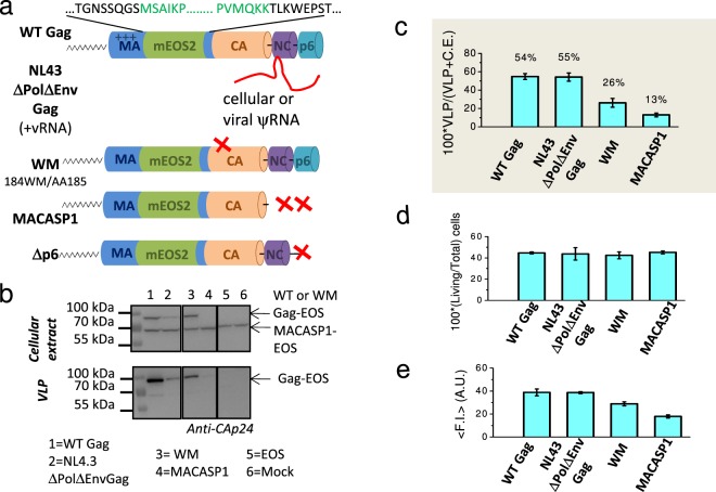
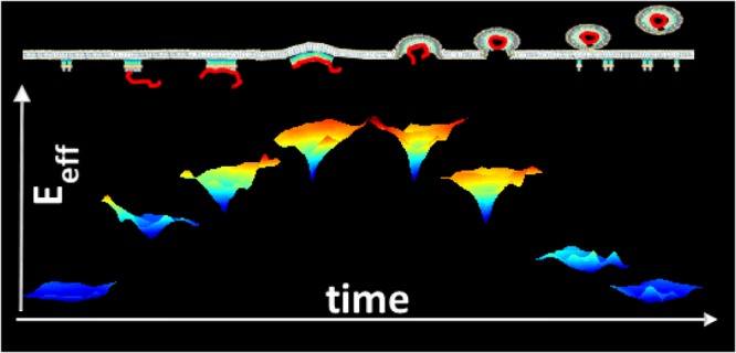
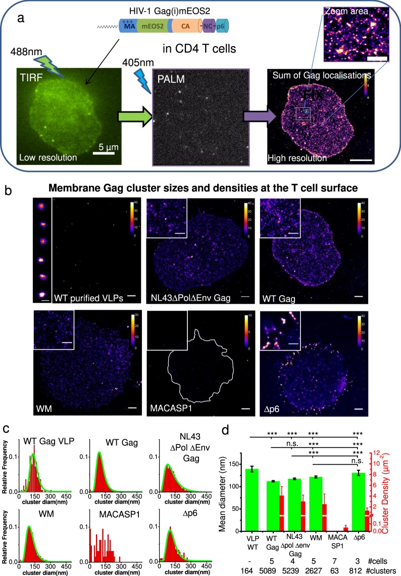
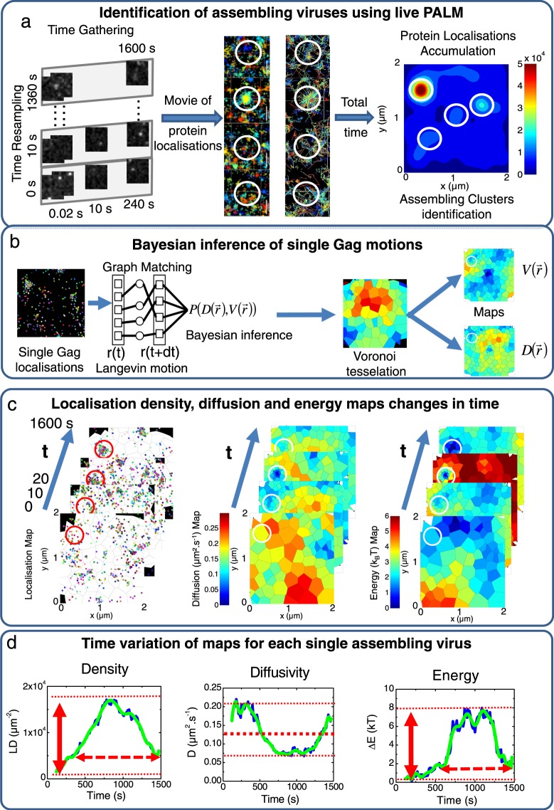
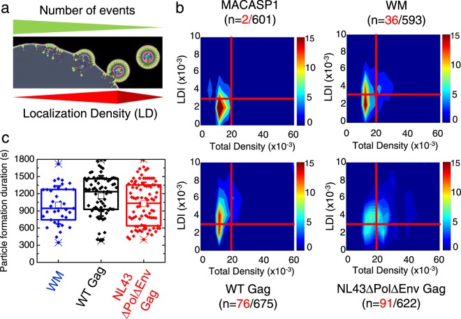
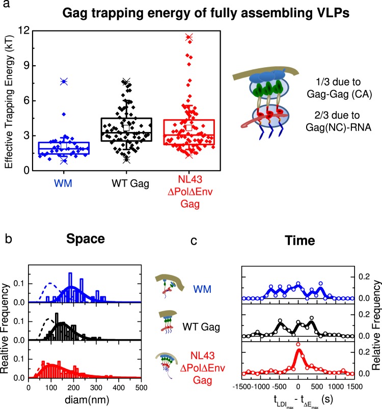
Monitoring virus assembly at the nanoscale in host cells remains a major challenge. Human immunodeficiency virus type 1 (HIV-1) components are addressed to the plasma membrane where they assemble to form spherical particles of 100 nm in diameter. Interestingly, HIV-1 Gag protein expression alone is sufficient to produce virus-like particles (VLPs) that resemble the immature virus. Here, we monitored VLP formation at the plasma membrane of host CD4 T cells using a newly developed workflow allowing the analysis of long duration recordings of single-molecule Gag protein localisation and movement. Comparison of Gag assembling platforms in CD4 T cells expressing wild type or assembly-defective Gag mutant proteins showed that VLP formation lasts roughly 15 minutes with an assembly time of 5 minutes. Trapping energy maps, built from membrane associated Gag protein movements, showed that one third of the assembling energy is due to direct Gag capsid-capsid interaction while the remaining two thirds require the nucleocapsid-RNA interactions. Finally, we show that the viral RNA genome does not increase the attraction of Gag at the membrane towards the assembling site but rather acts as a spatiotemporal coordinator of the membrane assembly process.
在宿主细胞中对纳米尺度下的病毒组装进行监测仍然是一个重大挑战。人类免疫缺陷病毒 1 型(HIV-1)的组成部分被递送到质膜,在那里它们组装成直径为 100nm 的球形颗粒。有趣的是,单独表达 HIV-1 Gag 蛋白就足以产生类似于不成熟病毒的病毒样颗粒(VLPs)。在这里,我们使用新开发的工作流程在宿主 CD4 T 细胞的质膜上监测 VLP 的形成,该流程允许对单个 Gag 蛋白定位和运动的长时间记录进行分析。比较在表达野生型或组装缺陷型 Gag 突变蛋白的 CD4 T 细胞中 Gag 组装平台的结果表明,VLP 的形成大约持续 15 分钟,组装时间为 5 分钟。从与膜相关的 Gag 蛋白运动构建的捕获能量图谱显示,组装能量的三分之一归因于直接 Gag 衣壳-衣壳相互作用,而剩余的三分之二则需要核衣壳-RNA 相互作用。最后,我们表明病毒 RNA 基因组不会增加 Gag 在膜上向组装部位的吸引力,而是作为膜组装过程的时空协调者。