Departments of Obstetrics and Gynecology, Osaka University Graduate School of Medicine, 2-2 Yamadaoka, Suita, Osaka, 565-0871, Japan.
Department of Molecular Oncology, H. Lee Moffitt Cancer Center & Research Institute, Tampa, FL, USA.
BMC Cancer. 2018 Nov 5;18(1):1065. doi: 10.1186/s12885-018-4974-5.
microRNAs (miRNAs) stably exist in circulating blood and are encapsulated in extracellular vesicles such as exosomes. The aims of this study were to identify which exosomal miRNAs are highly produced from epithelial ovarian cancer (EOC) cells, to analyze whether serum miRNA can be used to discriminate patients with EOC from healthy volunteers, and to investigate the functional role of exosomal miRNAs in ovarian cancer progression.
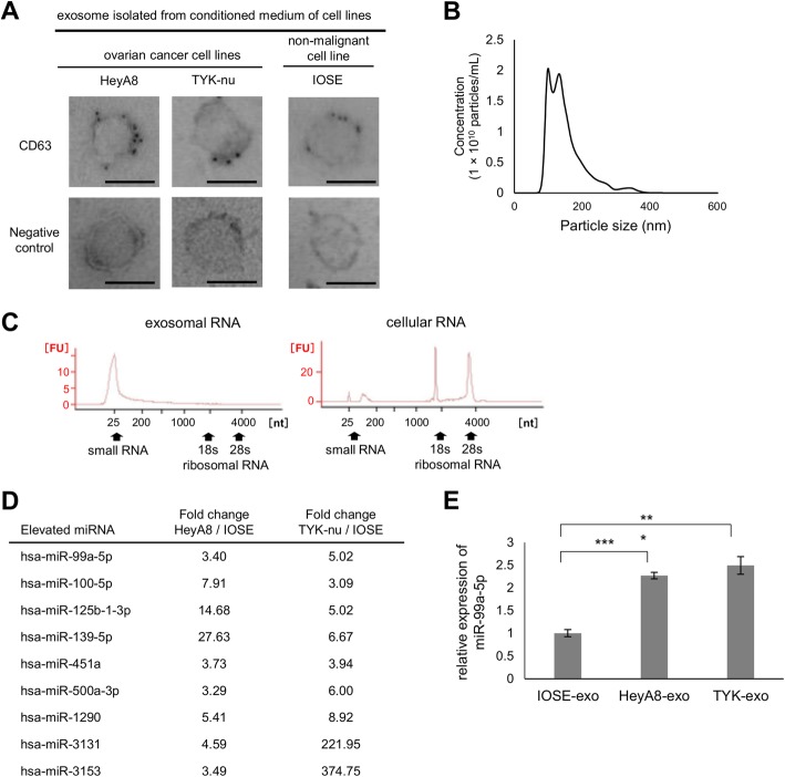
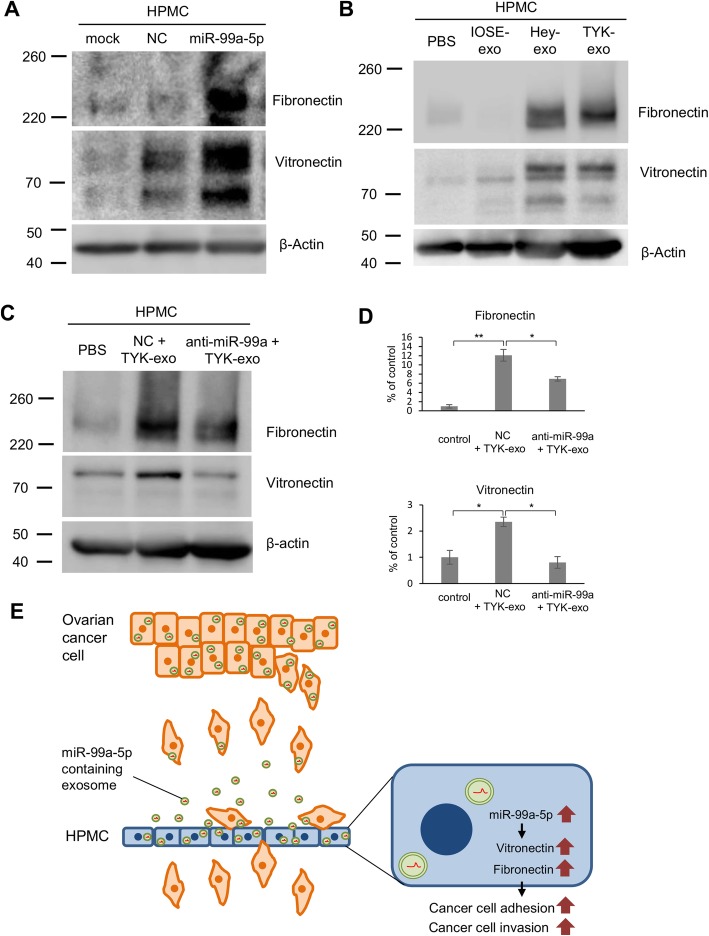
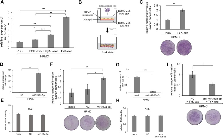
Exosomes were collected from the culture media of serous ovarian cancer cell lines, namely TYK-nu and HeyA8 cells. An exosomal miRNA microarray revealed that several miRNAs including miR-99a-5p were specifically elevated in EOC-derived exosomes. Expression levels of serum miR-99a-5p in 62 patients with EOC, 26 patients with benign ovarian tumors, and 20 healthy volunteers were determined by miRNA quantitative reverse transcription-polymerase chain reaction. To investigate the role of exosomal miR-99a-5p in peritoneal dissemination, neighboring human peritoneal mesothelial cells (HPMCs) were treated with EOC-derived exosomes and then expression levels of miR-99a-5p were examined. Furthermore, mimics of miR-99a-5p were transfected into HPMCs and the effect of miR-99a-5p on cancer invasion was analyzed using a 3D culture model. Proteomic analysis with the tandem mass tag method was performed on HPMCs transfected with miR-99a-5p and then potential target genes of miR-99a-5p were examined.
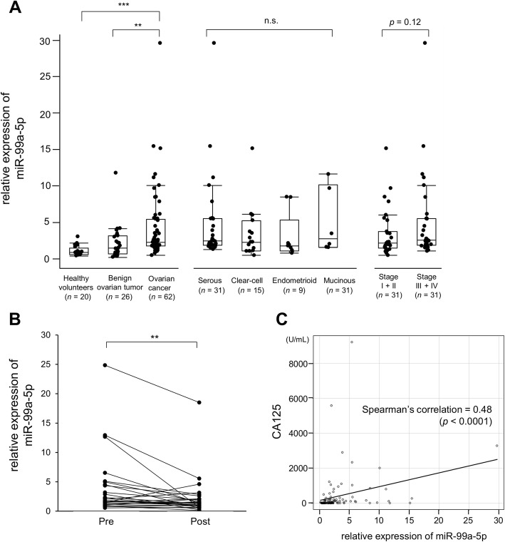
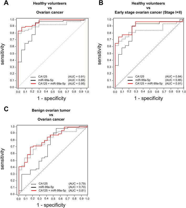
The serum miR-99a-5p levels were significantly increased in patients with EOC, compared with those in benign tumor patients and healthy volunteers (1.7-fold and 2.8-fold, respectively). A receiver operating characteristic curve analysis showed with a cut-off of 1.41 showed sensitivity and specificity of 0.85 and 0.75, respectively, for detecting EOC (area under the curve = 0.88). Serum miR-99a-5p expression levels were significantly decreased after EOC surgeries (1.8 to 1.3, p = 0.002), indicating that miR-99a-5p reflects tumor burden. Treatment with EOC-derived exosomes significantly increased miR-99a-5p expression in HPMCs. HPMCs transfected with miR-99a-5p promoted ovarian cancer invasion and exhibited increased expression levels of fibronectin and vitronectin.
Serum miR-99a-5p is significantly elevated in ovarian cancer patients. Exosomal miR-99a-5p from EOC cells promotes cell invasion by affecting HPMCs through fibronectin and vitronectin upregulation and may serve as a target for inhibiting ovarian cancer progression.
microRNAs (miRNAs) 稳定存在于循环血液中,并被包裹在细胞外囊泡中,如外泌体。本研究旨在鉴定哪些外泌体 miRNA 是由上皮性卵巢癌 (EOC) 细胞大量产生的,分析血清 miRNA 是否可用于区分 EOC 患者与健康志愿者,以及研究外泌体 miRNA 在卵巢癌进展中的功能作用。
从浆液性卵巢癌细胞系 TYK-nu 和 HeyA8 细胞的培养基中收集外泌体。外泌体 miRNA 微阵列显示,几种 miRNA(包括 miR-99a-5p)在上皮性卵巢癌来源的外泌体中特异性升高。通过 miRNA 定量逆转录聚合酶链反应测定 62 名 EOC 患者、26 名良性卵巢肿瘤患者和 20 名健康志愿者的血清 miR-99a-5p 表达水平。为了研究外泌体 miR-99a-5p 在腹膜扩散中的作用,用 EOC 来源的外泌体处理邻近的人腹膜间皮细胞 (HPMC),然后检测 miR-99a-5p 的表达水平。此外,将 miR-99a-5p 的模拟物转染到 HPMC 中,并使用 3D 培养模型分析 miR-99a-5p 对癌症侵袭的影响。用串联质量标签法对转染 miR-99a-5p 的 HPMC 进行蛋白质组学分析,然后检测 miR-99a-5p 的潜在靶基因。
与良性肿瘤患者和健康志愿者相比,EOC 患者的血清 miR-99a-5p 水平显著升高(分别为 1.7 倍和 2.8 倍)。受试者工作特征曲线分析显示,以 1.41 为截断值,检测 EOC 的敏感性和特异性分别为 0.85 和 0.75(曲线下面积为 0.88)。EOC 手术后血清 miR-99a-5p 表达水平显著降低(1.8 至 1.3,p=0.002),表明 miR-99a-5p 反映肿瘤负荷。用 EOC 来源的外泌体处理可显著增加 HPMC 中 miR-99a-5p 的表达。转染 miR-99a-5p 的 HPMC 促进卵巢癌细胞侵袭,并表现出纤维连接蛋白和 vitronectin 表达水平升高。
卵巢癌患者的血清 miR-99a-5p 水平显著升高。EOC 细胞来源的外泌体 miR-99a-5p 通过影响 HPMC 中的纤维连接蛋白和 vitronectin 而上调,促进细胞侵袭,可能成为抑制卵巢癌进展的靶点。