Dartmouth College, Thayer School of Engineering, Hanover, New Hampshire, United States.
Xidian University, Engineering Research Center of Molecular and Neuro Imaging of the Ministry of Edu, China.
J Biomed Opt. 2018 Nov;24(5):1-4. doi: 10.1117/1.JBO.24.5.051405.
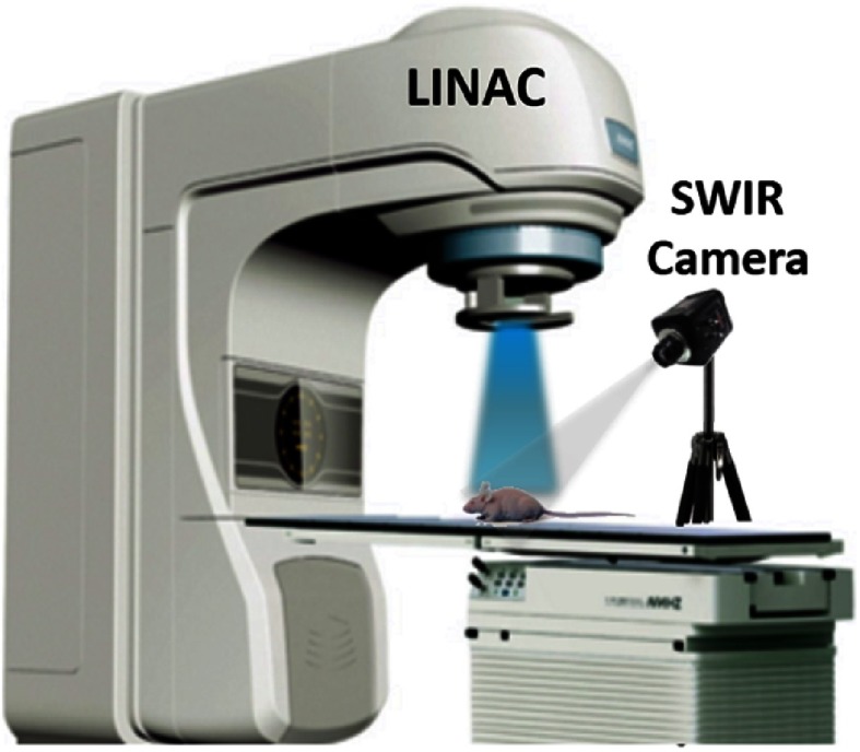
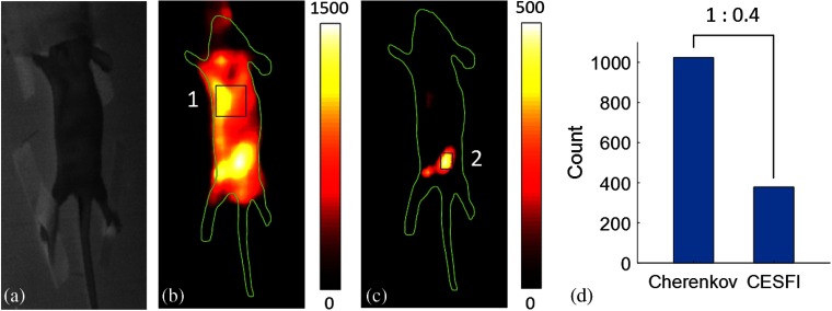
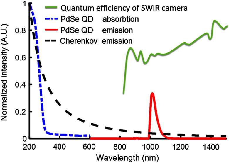
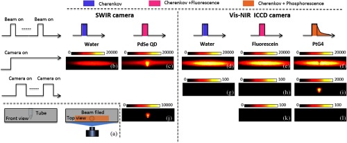
Cherenkov emission induced by external beam radiation therapy from a clinical linear accelerator (LINAC) can be used to excite phosphors deep in biological tissues. As with all luminescence imaging, there is a desire to minimize the spectral overlap between the excitation light and emission wavelengths, here between the Cherenkov and the phosphor. Cherenkov excited short-wavelength infrared (SWIR, 1000 to 1700 nm) fluorescence imaging has been demonstrated for the first time, using long Stokes-shift fluorophore PdSe quantum dots (QD) with nanosecond lifetime and an optimized SWIR detection. The 1 / λ2 intensity spectrum characteristic of Cherenkov emission leads to low overlap of this into the fluorescence spectrum of PdSe QDs in the SWIR range. Additionally, using a SWIR camera itself inherently ignores the stronger Cherenkov emission wavelengths dominant across the visible spectrum. The SWIR luminescence was shown to extend the depth sensitivity of Cherenkov imaging, which could be used for applications in radiotherapy sensing and imaging in human tissue with targeted molecular probes.
Cherenkov 辐射由临床线性加速器(LINAC)的外部射束辐射引发,可用于激发生物组织深处的荧光粉。与所有发光成像一样,人们希望最大限度地减少激发光与发射波长之间的光谱重叠,这里是 Cherenkov 辐射与荧光粉之间的光谱重叠。首次展示了使用具有纳秒寿命和优化的 SWIR 检测的长斯托克斯位移荧光团 PdSe 量子点(QD)进行的 Cherenkov 激发短波长红外(SWIR,1000 到 1700nm)荧光成像。Cherenkov 发射的 1 / λ2强度光谱特征导致其在 SWIR 范围内与 PdSe QD 的荧光光谱的低重叠。此外,使用 SWIR 相机本身就忽略了在可见光谱中占主导地位的更强的 Cherenkov 发射波长。SWIR 发光扩展了 Cherenkov 成像的深度灵敏度,可用于在人体组织中进行放射治疗传感和靶向分子探针成像的应用。