SDSU Heart Research Institute, San Diego State University, 5500 Campanile Drive, San Diego, CA 92182, USA.
SDSU Department of Mathematic and Statistics, San Diego State University, 5500 Campanile Drive, San Diego, CA 92182, USA.
J Mol Cell Cardiol. 2019 Feb;127:154-164. doi: 10.1016/j.yjmcc.2018.12.007. Epub 2018 Dec 18.
Understanding and manipulating the cardiomyocyte cell cycle has been the focus of decades of research, however the ultimate goal of activating mitotic activity in adult mammalian cardiomyocytes remains elusive and controversial. The relentless pursuit of controlling cardiomyocyte mitosis has been complicated and obfuscated by a multitude of indices used as evidence of cardiomyocyte cell cycle activity that lack clear identification of cardiomyocyte "proliferation" versus cell cycle progression, endoreplication, endomitosis, and even DNA damage. Unambiguous appreciation of the complexity of cardiomyocyte replication that avoids oversimplification and misinterpretation is desperately needed.
Track cardiomyocyte cell cycle activity and authenticate fidelity of proliferation markers as indicators of de novo cardiomyogenesis in post-mitotic cardiomyocytes.
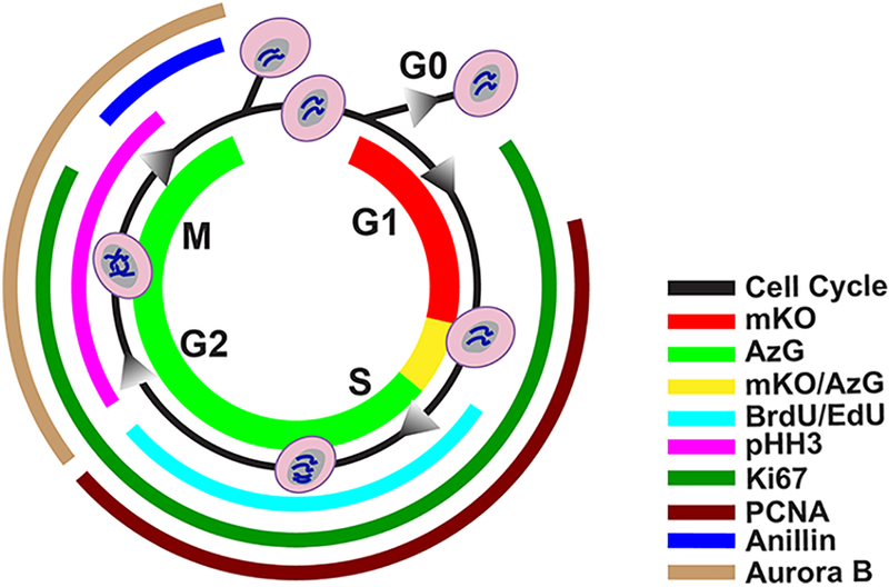
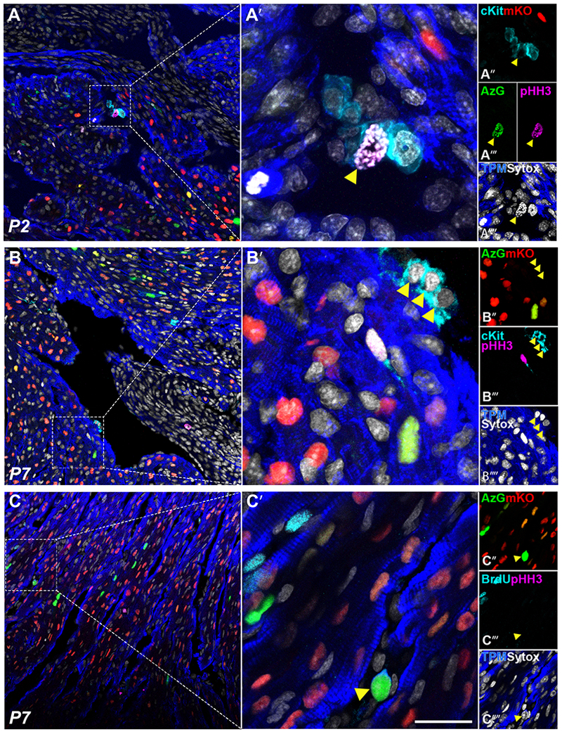
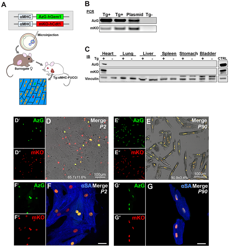
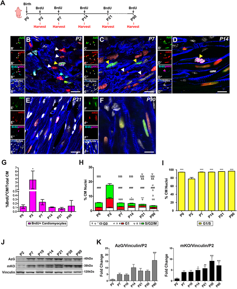
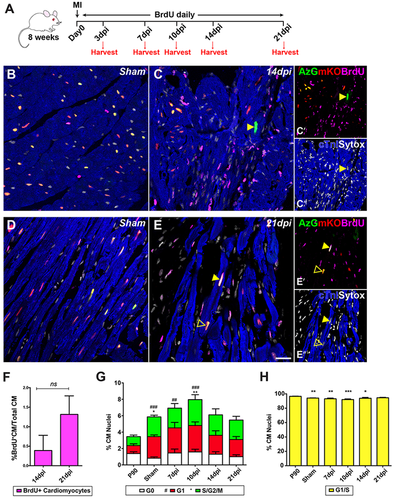
Cardiomyocytes expressing the FUCCI construct driven by the α-myosin heavy chain promoter were readily and uniformly detected through the myocardium of transgenic mice. Cardiomyocyte cell cycle activity peaks at postnatal day 2 and rapidly declines thereafter with almost all cardiomyocytes arrested at the G1/S cell cycle transition. Myocardial infarction injury in adult hearts prompts transient small increases in myocytes progressing through cell cycle without concurrent mitotic activity, indicating lack of cardiomyogenesis. In comparison, cardiomyogenic activity during early postnatal development correlated with coincidence of FUCCI and cKit cells that were undetectable in the adult myocardium.
Cardiomyocyte-specific expression of Fluorescence Ubiquitination-based Cell Cycle Indicators (FUCCI) reveals previously unappreciated aspects of cardiomyocyte cell cycle arrest and biological activity in postnatal development and in response to pathologic damage. Compared to many other methods and model systems, the FUCCI transgenic (FUCCI-Tg) mouse represents a valuable tool to unambiguously track cell cycle and proliferation of the entire cardiomyocyte population in the adult murine heart. FUCCI-Tg provides a desperately needed novel approach in the armamentarium of tools to validate cardiomyocyte proliferative activity that will reveal cell cycle progression, discriminate between cycle progression, DNA replication, and proliferation, and provide important insight for enhancing cardiomyocyte proliferation in the context of adult myocardial tissue.
几十年来,人们一直致力于理解和调控心肌细胞的细胞周期,然而,在成年哺乳动物心肌细胞中激活有丝分裂活性这一最终目标仍然难以捉摸且颇具争议。由于缺乏对心肌细胞“增殖”与细胞周期进程、核内复制、核内有丝分裂和 DNA 损伤的明确区分,因此,有丝分裂活动的控制这一目标一直被许多作为心肌细胞周期活性证据的指标所复杂化和混淆。明确认识心肌细胞复制的复杂性,避免过度简化和误解是当务之急。
追踪心肌细胞的细胞周期活动,并验证增殖标志物作为有丝分裂后心肌细胞新生的准确性。
通过转基因小鼠的心肌,很容易且均匀地检测到由α-肌球蛋白重链启动子驱动的 FUCCI 构建体表达的心肌细胞。心肌细胞的细胞周期活性在出生后第 2 天达到峰值,随后迅速下降,几乎所有的心肌细胞都在 G1/S 细胞周期转换处停滞。成年心脏的心肌梗死损伤会短暂地增加通过细胞周期的心肌细胞数量,但没有伴随有丝分裂活性,表明没有心肌发生。相比之下,在早期出生后发育过程中的心肌发生活性与 FUCCI 和 cKit 细胞的同时出现相关,而这些细胞在成年心肌中无法检测到。
荧光泛素化细胞周期指示剂(FUCCI)的心肌细胞特异性表达揭示了出生后发育过程中和对病理损伤反应中心肌细胞细胞周期阻滞和生物学活性的先前未被认识的方面。与许多其他方法和模型系统相比,FUCCI 转基因(FUCCI-Tg)小鼠是一种非常有价值的工具,可明确追踪成年鼠心脏中整个心肌细胞群体的细胞周期和增殖。FUCCI-Tg 为验证心肌细胞增殖活性提供了一种急需的新方法,该方法将揭示细胞周期进展,区分周期进展、DNA 复制和增殖,并为增强成年心肌组织中的心肌细胞增殖提供重要的见解。