• 文献检索
  • 文档翻译
  • 深度研究
  • 学术资讯
  • Suppr Zotero 插件Zotero 插件
  • 邀请有礼
  • 套餐&价格
  • 历史记录
应用&插件
Suppr Zotero 插件Zotero 插件浏览器插件Mac 客户端Windows 客户端微信小程序
定价
高级版会员购买积分包购买API积分包
服务
文献检索文档翻译深度研究API 文档MCP 服务
关于我们
关于 Suppr公司介绍联系我们用户协议隐私条款
关注我们

Suppr 超能文献

核心技术专利:CN118964589B侵权必究
粤ICP备2023148730 号-1Suppr @ 2026

文献检索

告别复杂PubMed语法,用中文像聊天一样搜索,搜遍4000万医学文献。AI智能推荐,让科研检索更轻松。

立即免费搜索

文件翻译

保留排版,准确专业,支持PDF/Word/PPT等文件格式,支持 12+语言互译。

免费翻译文档

深度研究

AI帮你快速写综述,25分钟生成高质量综述,智能提取关键信息,辅助科研写作。

立即免费体验

用于电子衍射的低温聚焦离子束蛋白质微晶制备

protein micro-crystal fabrication by cryo-FIB for electron diffraction.

作者信息

Li Xinmei, Zhang Shuangbo, Zhang Jianguo, Sun Fei

机构信息

1National Key Laboratory of Biomacromolecules, CAS Center for Excellence in Biomacromolecules, Institute of Biophysics, Chinese Academy of Sciences, Beijing, 100101 China.

2University of Chinese Academy of Sciences, Beijing, 100049 China.

出版信息

Biophys Rep. 2018;4(6):339-347. doi: 10.1007/s41048-018-0075-x. Epub 2018 Nov 14.

DOI:10.1007/s41048-018-0075-x
PMID:30596142
原文链接:https://pmc.ncbi.nlm.nih.gov/articles/PMC6276065/
Abstract

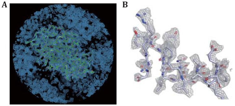
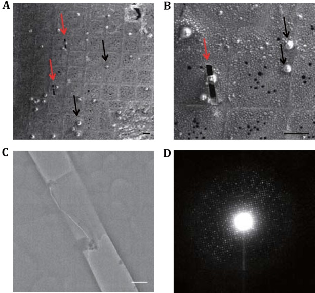
Micro-electron diffraction (MicroED) is an emerging technique to use cryo-electron microscope to study the crystal structures of macromolecule from its micro-/nano-crystals, which are not suitable for conventional X-ray crystallography. However, this technique has been prevented for its wide application by the limited availability of producing good micro-/nano-crystals and the inappropriate transfer of crystals. Here, we developed a complete workflow to prepare suitable crystals efficiently for MicroED experiment. This workflow includes on-grid crystallization, single-side blotting, cryo-focus ion beam (cryo-FIB) fabrication, and cryo-electron diffraction of crystal cryo-lamella. This workflow enables us to apply MicroED to study many small macromolecular crystals with the size of 2-10 μm, which is too large for MicroED but quite small for conventional X-ray crystallography. We have applied this method to solve 2.5 Å crystal structure of lysozyme from its micro-crystal within the size of 10 × 10 × 10 μm. Our work will greatly expand the availability space of crystals suitable for MicroED and fill up the gap between MicroED and X-ray crystallography.

摘要

微电子衍射(MicroED)是一种利用低温电子显微镜从微/纳米晶体研究大分子晶体结构的新兴技术,这些微/纳米晶体不适用于传统的X射线晶体学。然而,由于难以获得高质量的微/纳米晶体以及晶体转移不当,该技术的广泛应用受到了限制。在此,我们开发了一套完整的工作流程,能够高效地制备适合MicroED实验的晶体。该工作流程包括在网格上结晶、单面印迹、低温聚焦离子束(cryo-FIB)制备以及晶体低温薄片的低温电子衍射。这一工作流程使我们能够应用MicroED研究许多尺寸为2-10μm的小型大分子晶体,这些晶体对于MicroED来说太大,但对于传统X射线晶体学来说又太小。我们已应用此方法解析了尺寸为10×10×10μm的溶菌酶微晶的2.5Å晶体结构。我们的工作将极大地拓展适合MicroED的晶体的可得空间,并填补MicroED与X射线晶体学之间的空白。

https://cdn.ncbi.nlm.nih.gov/pmc/blobs/6452/6276065/df1a4199ba99/41048_2018_75_Fig5_HTML.jpg
https://cdn.ncbi.nlm.nih.gov/pmc/blobs/6452/6276065/086a999b45db/41048_2018_75_Fig1_HTML.jpg
https://cdn.ncbi.nlm.nih.gov/pmc/blobs/6452/6276065/652d589d4f60/41048_2018_75_Fig2_HTML.jpg
https://cdn.ncbi.nlm.nih.gov/pmc/blobs/6452/6276065/200e593e3677/41048_2018_75_Fig3_HTML.jpg
https://cdn.ncbi.nlm.nih.gov/pmc/blobs/6452/6276065/01f447bba89a/41048_2018_75_Fig4_HTML.jpg
https://cdn.ncbi.nlm.nih.gov/pmc/blobs/6452/6276065/df1a4199ba99/41048_2018_75_Fig5_HTML.jpg

相似文献

1
protein micro-crystal fabrication by cryo-FIB for electron diffraction.用于电子衍射的低温聚焦离子束蛋白质微晶制备
Biophys Rep. 2018;4(6):339-347. doi: 10.1007/s41048-018-0075-x. Epub 2018 Nov 14.
2
Using focus ion beam to prepare crystal lamella for electron diffraction.利用聚焦离子束制备电子衍射晶体薄片。
J Struct Biol. 2019 Mar 1;205(3):59-64. doi: 10.1016/j.jsb.2019.02.004. Epub 2019 Feb 20.
3
Collection of Continuous Rotation MicroED Data from Ion Beam-Milled Crystals of Any Size.从任何尺寸的离子束铣削晶体中收集连续旋转微 ED 数据。
Structure. 2019 Mar 5;27(3):545-548.e2. doi: 10.1016/j.str.2018.12.003. Epub 2019 Jan 17.
4
Studying Membrane Protein Structures by MicroED.利用微电镜断层扫描技术研究膜蛋白结构
Methods Mol Biol. 2021;2302:137-151. doi: 10.1007/978-1-0716-1394-8_8.
5
A Workflow for Protein Structure Determination From Thin Crystal Lamella by Micro-Electron Diffraction.一种通过微电子衍射从薄晶体薄片确定蛋白质结构的工作流程。
Front Mol Biosci. 2020 Aug 4;7:179. doi: 10.3389/fmolb.2020.00179. eCollection 2020.
6
Protein and Small Molecule Structure Determination by the Cryo-EM Method MicroED.利用冷冻电镜(Cryo-EM)方法的微束电子衍射(MicroED)进行蛋白质和小分子结构测定。
Methods Mol Biol. 2021;2305:323-342. doi: 10.1007/978-1-0716-1406-8_16.
7
Structure determination of a DNA crystal by MicroED.通过微晶电子衍射法测定DNA晶体的结构。
Structure. 2023 Dec 7;31(12):1499-1503.e2. doi: 10.1016/j.str.2023.07.005. Epub 2023 Aug 3.
8
Qualitative Analyses of Polishing and Precoating FIB Milled Crystals for MicroED.用于微 ED 的 FIB 铣削晶体的抛光和预涂的定性分析。
Structure. 2019 Oct 1;27(10):1594-1600.e2. doi: 10.1016/j.str.2019.07.004. Epub 2019 Aug 15.
9
A Rare Lysozyme Crystal Form Solved Using Highly Redundant Multiple Electron Diffraction Datasets from Micron-Sized Crystals.一种罕见的溶菌酶晶体形式,使用来自微米级晶体的高度冗余的多个电子衍射数据集解决。
Structure. 2018 Apr 3;26(4):667-675.e3. doi: 10.1016/j.str.2018.02.015. Epub 2018 Mar 15.
10
Atomic resolution structure determination by the cryo-EM method MicroED.通过低温电子显微镜方法MicroED进行原子分辨率结构测定。
Protein Sci. 2017 Jan;26(1):8-15. doi: 10.1002/pro.2989. Epub 2016 Aug 19.

引用本文的文献

1
VMXm - A sub-micron focus macromolecular crystallography beamline at Diamond Light Source.VMXm——钻石光源的一条亚微米聚焦大分子晶体学光束线。
J Synchrotron Radiat. 2024 Nov 1;31(Pt 6):1593-1608. doi: 10.1107/S1600577524009160. Epub 2024 Oct 30.
2
Structure determination of a DNA crystal by MicroED.通过微晶电子衍射法测定DNA晶体的结构。
Structure. 2023 Dec 7;31(12):1499-1503.e2. doi: 10.1016/j.str.2023.07.005. Epub 2023 Aug 3.
3
Design and implementation of suspended drop crystallization.悬浮液滴结晶的设计与实现。

本文引用的文献

1
From electron crystallography of 2D crystals to MicroED of 3D crystals.从二维晶体的电子晶体学到三维晶体的微晶电子衍射。
Curr Opin Colloid Interface Sci. 2018 Mar;34:9-16. doi: 10.1016/j.cocis.2018.01.010. Epub 2018 Jan 31.
2
Atomic structures of FUS LC domain segments reveal bases for reversible amyloid fibril formation.FUS LC 结构域片段的原子结构揭示了其可逆淀粉样纤维形成的基础。
Nat Struct Mol Biol. 2018 Apr;25(4):341-346. doi: 10.1038/s41594-018-0050-8. Epub 2018 Apr 2.
3
Serial femtosecond crystallography at the SACLA: breakthrough to dynamic structural biology.
IUCrJ. 2023 Jul 1;10(Pt 4):430-436. doi: 10.1107/S2052252523004141.
4
MicroED for the study of protein-ligand interactions and the potential for drug discovery.用于蛋白质-配体相互作用研究及药物发现潜力的微晶电子衍射技术。
Nat Rev Chem. 2021 Dec;5(12):853-858. doi: 10.1038/s41570-021-00332-y. Epub 2021 Oct 27.
5
Design and implementation of suspended drop crystallization.悬滴结晶法的设计与实现
bioRxiv. 2023 Mar 28:2023.03.28.534639. doi: 10.1101/2023.03.28.534639.
6
Investigation of the milling characteristics of different focused-ion-beam sources assessed by three-dimensional electron diffraction from crystal lamellae.利用晶体薄片的三维电子衍射评估不同聚焦离子束源的铣削特性研究。
IUCrJ. 2023 May 1;10(Pt 3):270-287. doi: 10.1107/S2052252523001902.
7
A robust approach for MicroED sample preparation of lipidic cubic phase embedded membrane protein crystals.一种用于嵌入脂质立方相的膜蛋白晶体的 MicroED 样品制备的稳健方法。
Nat Commun. 2023 Feb 25;14(1):1086. doi: 10.1038/s41467-023-36733-4.
8
ELI trifocal microscope: a precise system to prepare target cryo-lamellae for in situ cryo-ET study.ELI 三焦点显微镜:用于原位冷冻电镜研究的目标冷冻薄片制备的精确系统。
Nat Methods. 2023 Feb;20(2):276-283. doi: 10.1038/s41592-022-01748-0. Epub 2023 Jan 16.
9
Ab initio phasing macromolecular structures using electron-counted MicroED data.利用电子计数 MicroED 数据进行从头解析大分子结构。
Nat Methods. 2022 Jun;19(6):724-729. doi: 10.1038/s41592-022-01485-4. Epub 2022 May 30.
10
MicroED: conception, practice and future opportunities.微电子衍射:概念、实践与未来机遇。
IUCrJ. 2022 Jan 18;9(Pt 2):169-179. doi: 10.1107/S2052252521013063. eCollection 2022 Mar 1.
日本兵库县的SPring-8Ångström自由电子激光装置的串行飞秒晶体学:向动态结构生物学的突破。
Biophys Rev. 2018 Apr;10(2):209-218. doi: 10.1007/s12551-017-0344-9. Epub 2017 Dec 1.
4
A Bright Future for Serial Femtosecond Crystallography with XFELs.X射线自由电子激光驱动的飞秒串列晶体学的光明未来。
Trends Biochem Sci. 2017 Sep;42(9):749-762. doi: 10.1016/j.tibs.2017.06.007. Epub 2017 Jul 18.
5
Light-induced structural changes and the site of O=O bond formation in PSII caught by XFEL.X 射线自由电子激光捕获 PSII 中光诱导结构变化和 O=O 键形成的位置。
Nature. 2017 Mar 2;543(7643):131-135. doi: 10.1038/nature21400. Epub 2017 Feb 20.
6
Ab initio structure determination from prion nanocrystals at atomic resolution by MicroED.通过微电子衍射从朊病毒纳米晶体中以原子分辨率从头确定结构。
Proc Natl Acad Sci U S A. 2016 Oct 4;113(40):11232-11236. doi: 10.1073/pnas.1606287113. Epub 2016 Sep 19.
7
Atomic resolution structure determination by the cryo-EM method MicroED.通过低温电子显微镜方法MicroED进行原子分辨率结构测定。
Protein Sci. 2017 Jan;26(1):8-15. doi: 10.1002/pro.2989. Epub 2016 Aug 19.
8
Analysis of chromatin fibers in Hela cells with electron tomography.利用电子断层扫描技术分析海拉细胞中的染色质纤维。
Biophys Rep. 2015;1:51-60. doi: 10.1007/s41048-015-0009-9. Epub 2015 Aug 7.
9
An improved cryo-FIB method for fabrication of frozen hydrated lamella.一种用于制备冷冻水合薄片的改进型冷冻聚焦离子束方法。
J Struct Biol. 2016 May;194(2):218-23. doi: 10.1016/j.jsb.2016.02.013. Epub 2016 Feb 12.
10
Simultaneous determination of sample thickness, tilt, and electron mean free path using tomographic tilt images based on Beer-Lambert law.基于比尔-朗伯定律,利用断层倾斜图像同时测定样品厚度、倾斜度和电子平均自由程。
J Struct Biol. 2015 Nov;192(2):287-96. doi: 10.1016/j.jsb.2015.09.019. Epub 2015 Oct 9.