Department of Bioengineering, University of Illinois at Urbana-Champaign, Urbana, Illinois, 61801, USA.
Quantitative Light Imaging Laboratory, Department of Electrical and Computer Engineering, Beckman Institute for Advanced Science and Technology, University of Illinois at Urbana-Champaign, Urbana, Illinois, 61801, USA.
Sci Rep. 2019 Jan 22;9(1):248. doi: 10.1038/s41598-018-36551-5.
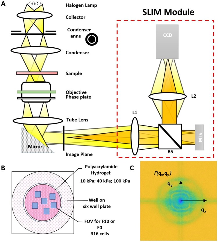
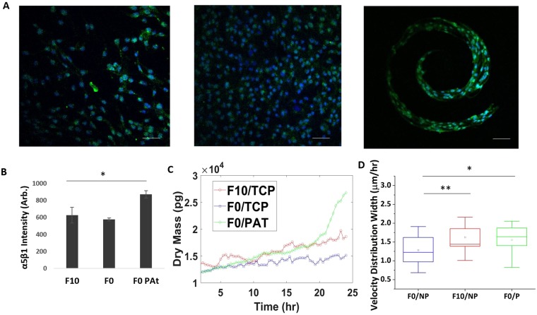
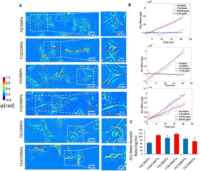
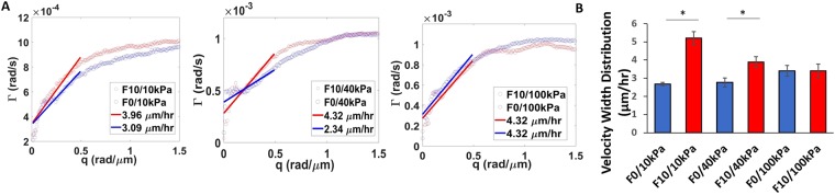
Cancer progression involves complex signals within the tumor microenvironment that orchestrate proliferation and invasive processes. The mechanical properties of the extracellular matrix (ECM) within this microenvironment has been demonstrated to influence growth and the migratory phenotype that precedes invasion. Here we present the integration of a label-free quantitative phase imaging technique, spatial light interference microscopy (SLIM)-with protein-conjugated hydrogel substrates-to explore how the stiffness of the ECM influences melanoma cells of varying metastatic potential. Melanoma cells of high metastatic potential demonstrate increased growth and velocity characteristics relative to cells of low metastatic potential. Cell velocity in the highly metastatic population shows a relative insensitivity to matrix stiffness suggesting adoption of migratory routines that are independent of mechanics to facilitate invasion. The use of SLIM and engineered substrates provides a new approach to characterize the invasive properties of live cells as a function of microenvironment parameters. This work provides fundamental insight into the relationship between growth, migration and metastatic potential, and provides a new tool for profiling cancer cells for clinical grading and development of patient-specific therapeutic regimens.
癌症的进展涉及肿瘤微环境中的复杂信号,这些信号协调增殖和侵袭过程。已经证明,细胞外基质(ECM)的机械性能会影响生长和侵袭前的迁移表型。在这里,我们介绍了一种无标记定量相位成像技术(空间光干涉显微镜,SLIM)与蛋白偶联水凝胶底物的整合,以探讨 ECM 的硬度如何影响具有不同转移潜能的黑色素瘤细胞。高转移潜能的黑色素瘤细胞相对于低转移潜能的细胞表现出更高的生长和速度特征。在高转移群体中,细胞速度对基质硬度的相对不敏感表明采用了独立于力学的迁移常规,以促进侵袭。SLIM 和工程化底物的使用为研究活细胞的侵袭特性提供了一种新方法,这些特性可作为微环境参数的函数进行描述。这项工作为生长、迁移和转移潜能之间的关系提供了基本的见解,并为用于临床分级和制定患者特异性治疗方案的癌症细胞分析提供了一种新工具。