Philipps University of Marburg, Dental School, Department of Orthodontics, Marburg, Germany.
MH Statistics Consulting, Marburg, Germany.
Sci Rep. 2019 Jan 22;9(1):269. doi: 10.1038/s41598-018-36536-4.
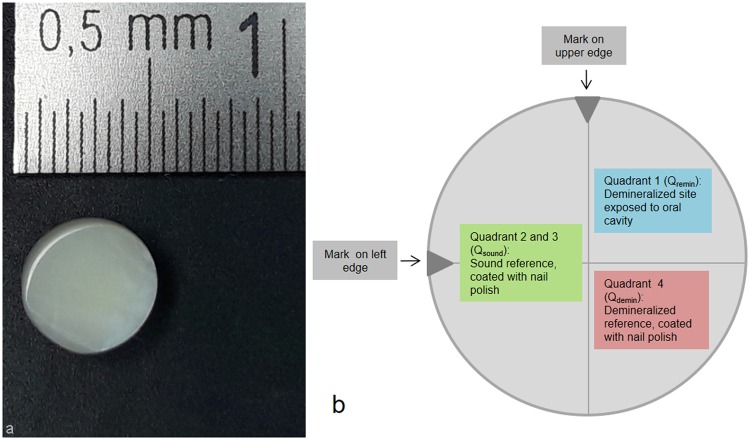
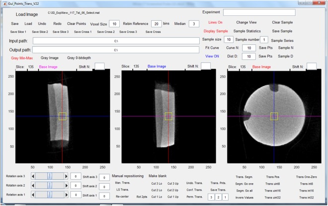
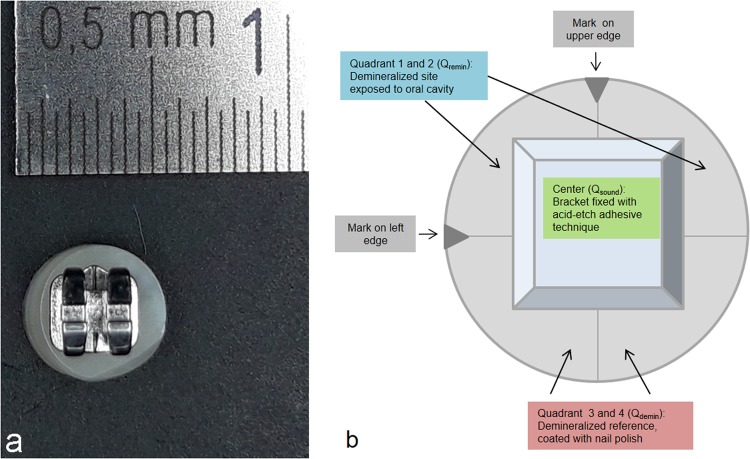
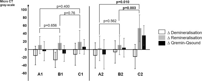
The aim was to investigate the ability of self-assembling Peptide P11-4 Matrix (SAPM) to remineralize artificial initial caries lesions compared to the use of fluoride varnish. Volunteers were recruited for this randomised, cross-over in situ trial. Bovine specimens, half including orthodontic brackets, were recessed on the buccal aspects of mandibular appliances. Specimens included internal sound enamel control, a demineralised control and a part exposed during the in situ phase. Each phase lasted four weeks, followed by a one-week washout. Treatment groups were: A: negative control, no treatment,B: positive control, 22,600 ppm fluoride varnish,C: test group, 1,000 ppm SAPM. Laser fluorescence values (LF) were measured before/after demineralisation, and after the in situ period. Micro-CT analysis was used to assess mineral changes within the specimens after the in situ phase. In specimens without brackets, ΔLF values after in situ phase were: A: +5.28, B: +0.85, C: -2.89. Corresponding ΔLF for specimens with brackets were: A: +5.77, B: +1.30, C: -3.15. LF-values between groups significantly differed from each other (p < 0.0001) after the in situ phase. Micro-CT analysis yielded no significant difference among groups for specimens without brackets. For specimens with brackets, the test group showed significantly more remineralisation than the negative (p = 0.01) and positive control (p = 0.003). Within the limitations of the study, SAPM showed prevention of caries and remineralisation of enamel around orthodontic brackets.
目的是研究自组装肽 P11-4 基质(SAPM)与氟化物漆相比,再矿化人工初始龋损的能力。志愿者被招募参加这项随机、交叉原位试验。牛标本,其中一半包括正畸托槽,被凹陷在下颌矫治器的颊面。标本包括内部健全的釉质对照、脱矿质对照和在原位阶段暴露的部分。每个阶段持续四周,然后进行一周的洗脱。治疗组为:A:阴性对照,无治疗,B:阳性对照,22600ppm 氟化物漆,C:试验组,1000ppm SAPM。在脱矿质前后以及原位阶段后测量激光荧光值(LF)。微 CT 分析用于评估原位阶段后标本内的矿物质变化。在没有托槽的标本中,原位阶段后ΔLF 值为:A:+5.28,B:+0.85,C:-2.89。相应的带托槽标本的ΔLF 值为:A:+5.77,B:+1.30,C:-3.15。原位阶段后,各组 LF 值之间存在显著差异(p<0.0001)。对于没有托槽的标本,微 CT 分析结果在各组之间没有显著差异。对于有托槽的标本,试验组的再矿化程度明显高于阴性组(p=0.01)和阳性对照组(p=0.003)。在研究的限制范围内,SAPM 显示出对龋齿的预防和正畸托槽周围釉质的再矿化作用。