IEEE Trans Med Imaging. 2019 Aug;38(8):1812-1820. doi: 10.1109/TMI.2019.2897044. Epub 2019 Feb 1.
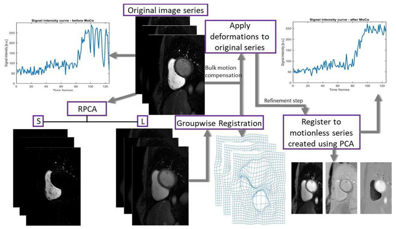
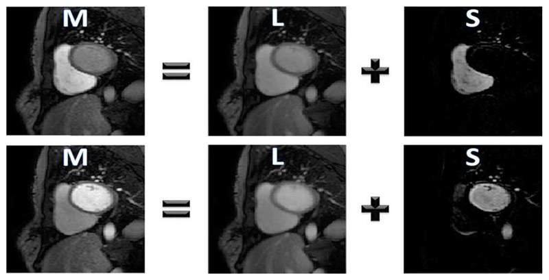
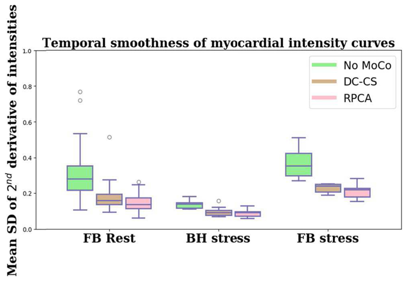
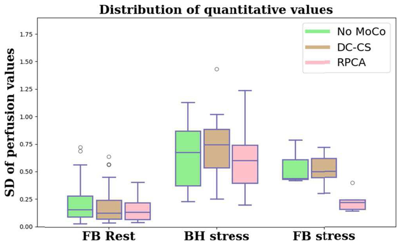
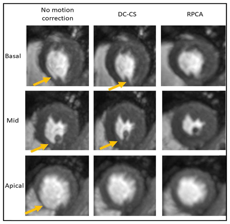
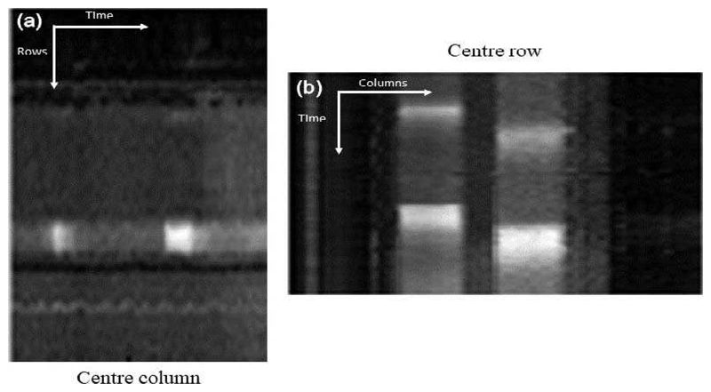
Kinetic parameter values, such as myocardial perfusion, can be quantified from dynamic contrast-enhanced magnetic resonance imaging data using tracer-kinetic modeling. However, respiratory motion affects the accuracy of this process. Motion compensation of the image series is difficult due to the rapid local signal enhancement caused by the passing of the gadolinium-based contrast agent. This contrast enhancement invalidates the assumptions of the (global) cost functions traditionally used in intensity-based registrations. The algorithms are unable to distinguish whether the differences in signal intensity between frames are caused by the spatial motion artifacts or the local contrast enhancement. In order to address this problem, a fully automated motion compensation scheme is proposed, which consists of two stages. The first of which uses robust principal component analysis (PCA) to separate the local signal enhancement from the baseline signal, before a refinement stage which uses the traditional PCA to construct a synthetic reference series that is free from motion but preserves the signal enhancement. Validation is performed on 18 subjects acquired in free-breathing and 5 clinical subjects acquired with a breath-hold. The validation assesses the visual quality, the temporal smoothness of tissue curves, and the clinically relevant quantitative perfusion values. The expert observers score the visual quality increased by a mean of 1.58/5 after motion compensation and improvement over the previously published methods. The proposed motion compensation scheme also leads to the improved quantitative performance of motion compensated free-breathing image series [30% reduction in the coefficient of variation across quantitative perfusion maps and 53% reduction in temporal variations (p < 0.001)].
动力学参数值,如心肌灌注,可以从动态对比增强磁共振成像数据中使用示踪动力学建模来定量。然而,呼吸运动影响这个过程的准确性。由于钆基造影剂的通过导致局部信号快速增强,图像序列的运动补偿是困难的。这种对比增强使传统基于强度的配准中使用的(全局)成本函数的假设无效。算法无法区分帧间信号强度的差异是由空间运动伪影还是局部对比增强引起的。为了解决这个问题,提出了一种完全自动化的运动补偿方案,该方案由两个阶段组成。第一阶段使用稳健主成分分析(PCA)将局部信号增强与基线信号分离,然后在精炼阶段使用传统 PCA 构建无运动但保留信号增强的合成参考序列。在自由呼吸采集的 18 个受试者和呼吸暂停采集的 5 个临床受试者上进行验证。验证评估了视觉质量、组织曲线的时间平滑度和临床相关的定量灌注值。专家观察者对视觉质量的评分平均提高了 1.58/5,并且优于之前发表的方法。所提出的运动补偿方案还提高了运动补偿自由呼吸图像序列的定量性能[定量灌注图的变异系数降低 30%,时间变化降低 53%(p < 0.001)]。