Department of Radiology, Mayo Clinic College of Medicine and Science, Rochester, Minnesota, USA.
Department of Physiology and Biomedical Engineering, Mayo Clinic College of Medicine and Science, Rochester, Minnesota, USA.
Ultrasound Med Biol. 2019 Apr;45(4):1010-1018. doi: 10.1016/j.ultrasmedbio.2018.10.028. Epub 2019 Feb 2.
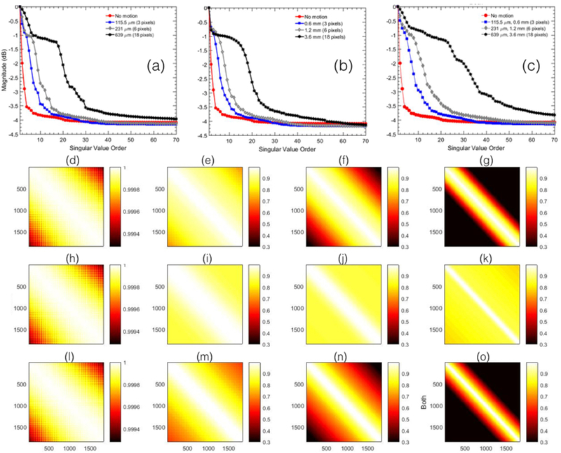
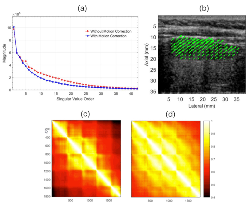
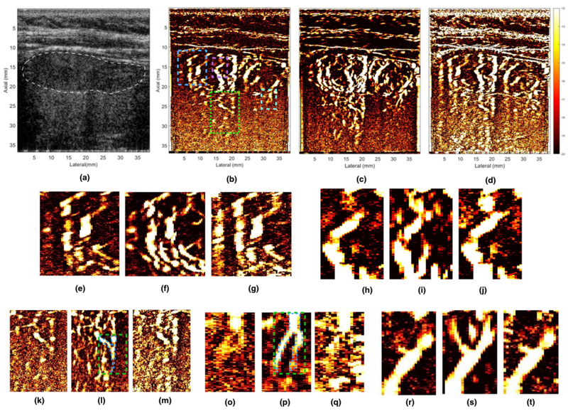
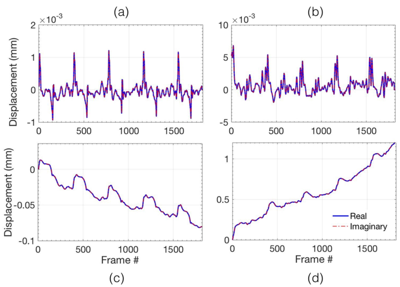
Reliable assessment of small vessel blood flow in the thyroid, without using any contrast agents, can be challenging because of increased physiological motion resulting from its proximity to the pulsating carotid artery. In this study, we hypothesized that correction of tissue motion prior to singular value decomposition (SVD)-based clutter filtering can improve the coherency of the tissue components and, thus, may allow better clutter suppression and visualization of small vessels in the thyroid. We corroborated this hypothesis by conducting phantom and in vivo studies using a clinical ultrasound scanner implemented with compounded plane wave imaging. The phantom studies were conducted using a homogeneous tissue-mimicking phantom to study the impact of motion on the covariance of the spatiotemporal Doppler data, in the absence of blood activity. The non-invasive in vivo study was conducted on a 74-y-old woman with a thyroid nodule suspicious of malignancy. A rigid body-based motion correction was performed using tissue displacements obtained from 2-D normalized cross-correlation-based speckle tracking. Subsequently, the power Doppler images were computed using SVD-based spatiotemporal clutter filtering. The results from the phantom study revealed that motion can considerably reduce the covariance of the spatiotemporal data and, thus, increase the rank of the tissue components. When the phantom was subjected to a total translation displacement of 6 pixels over the entire ensemble, in each direction (axial and lateral), the covariance dropped by more than 25%. The results obtained from the non-invasive in vivo study indicated that visualization of small vessel blood flow improved with motion correction of the power Doppler ensemble. The contrast-to-noise ratio of the blood signal in motion-corrected power Doppler images was considerably higher (8.17 and 8.32 dB), compared with that obtained using the standard SVD approach at an optimal threshold (0.87 and 4.33 dB) and a lower singular value threshold (1.92 and 3.05 dB). Further, the covariance of the in vivo thyroid spatiotemporal data increased by approximately 10% with motion correction. These preliminary results indicate that motion correction can be used to improve the visualization of small vessel blood flow in the thyroid, without using any contrast agents. The results of this feasibility study were encouraging, and warrant further development and more in vivo validation in moving tissues and organs.
可靠地评估甲状腺中的小血管血流,而不使用任何造影剂,可能具有挑战性,因为其靠近脉动的颈动脉,导致生理运动增加。在这项研究中,我们假设在基于奇异值分解(SVD)的杂波滤波之前对组织运动进行校正,可以改善组织成分的相干性,从而可以更好地抑制杂波并可视化甲状腺中的小血管。我们通过使用配备复合平面波成像的临床超声扫描仪进行的体模和体内研究证实了这一假设。体模研究使用均匀的组织模拟体模进行,以研究运动对时空多普勒数据协方差的影响,而不考虑血液活动。非侵入性的体内研究是在一位 74 岁的女性身上进行的,她患有甲状腺结节,疑似恶性肿瘤。使用基于 2D 归一化互相关斑点跟踪的组织位移进行了基于刚体的运动校正。随后,使用基于 SVD 的时空杂波滤波计算功率多普勒图像。体模研究的结果表明,运动可以大大降低时空数据的协方差,从而增加组织成分的秩。当整个集合中的每个方向(轴向和横向)的总平移位移为 6 像素时,协方差降低了 25%以上。非侵入性体内研究的结果表明,运动校正后,小血管血流的可视化得到改善。运动校正后的功率多普勒图像中的血流信号的对比噪声比明显更高(8.17 和 8.32dB),与使用标准 SVD 方法在最佳阈值(0.87 和 4.33dB)和较低奇异值阈值(1.92 和 3.05dB)时获得的结果相比。此外,运动校正后,体内甲状腺时空数据的协方差增加了约 10%。这些初步结果表明,运动校正可用于改善甲状腺中无需使用任何造影剂的小血管血流可视化。这项可行性研究的结果令人鼓舞,需要在移动组织和器官中进一步开发和更多的体内验证。