Department of Psychiatry, University of Toronto, Toronto, ON, Canada.
Clinical Research Services, The Hospital for Sick Children, and the Dalla Lana School of Public Health, University of Toronto, Toronto, ON, Canada.
Transl Psychiatry. 2019 Feb 4;9(1):72. doi: 10.1038/s41398-019-0382-0.
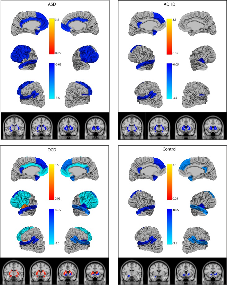
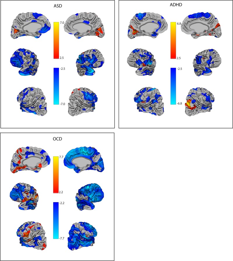
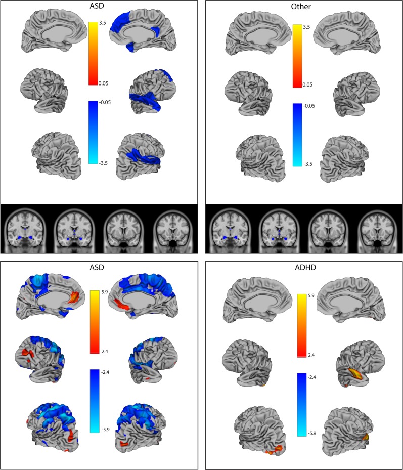
Autism spectrum disorder (ASD), attention-deficit/hyperactivity disorder (ADHD), and obsessive-compulsive disorder (OCD) have been associated with difficulties recognizing and responding to social cues. Neuroimaging studies have begun to map the social brain; however, the specific neural substrates contributing to social deficits in neurodevelopmental disorders remain unclear. Three hundred and twelve children underwent structural magnetic resonance imaging of the brain (controls = 32, OCD = 44, ADHD = 77, ASD = 159; mean age = 11). Their social deficits were quantified on the Social Communication Questionnaire (SCQ) and the Reading the Mind in the Eyes Test (RMET). Multivariable regression models were used to examine the structural neuroimaging correlates of social deficits, with both a region of interest and a whole-brain vertex-wise approach. For the region of interest analysis, social brain regions were grouped into three networks: (1) lateral mentalization (e.g., temporal-parietal junction), (2) frontal cognitive (e.g., orbitofrontal cortex), and (3) subcortical affective (e.g., limbic system) regions. Overall, social communication deficits on the SCQ were associated with thinner cortices in the left lateral regions and the right insula, and decreased volume in the ventral striatum, across diagnostic groups (p = 0.006 to <0.0001). Smaller subcortical volumes were associated with more severe social deficits on the SCQ in ASD and ADHD, and less severe deficits in OCD. On the RMET, larger amygdala/hippocampal volumes were associated with fewer deficits across groups. Overall, patterns of associations were similar in ASD and ADHD, supporting a common underlying biology and the blurring of the diagnostic boundaries between these disorders.
自闭症谱系障碍 (ASD)、注意力缺陷多动障碍 (ADHD) 和强迫症 (OCD) 与识别和响应社交线索的困难有关。神经影像学研究已经开始绘制社交大脑图谱;然而,导致神经发育障碍中社交缺陷的特定神经基质仍不清楚。312 名儿童接受了大脑结构磁共振成像检查(对照组 = 32 名,OCD = 44 名,ADHD = 77 名,ASD = 159 名;平均年龄 = 11 岁)。他们的社交缺陷通过社交沟通问卷 (SCQ) 和读心测试 (RMET) 进行量化。多变量回归模型用于检查社会缺陷的结构神经影像学相关性,采用了感兴趣区域和全脑顶点方法。对于感兴趣区域分析,社交大脑区域被分为三个网络:(1) 侧心理化 (例如颞顶联合区),(2) 额认知 (例如眶额皮层),和 (3) 皮质下情感 (例如边缘系统) 区域。总的来说,SCQ 的社交沟通缺陷与左侧外侧区域和右侧脑岛的皮质变薄以及腹侧纹状体体积减少有关,在所有诊断组中(p = 0.006 至 <0.0001)。较小的皮质下体积与 ASD 和 ADHD 中 SCQ 的更严重社交缺陷以及 OCD 中更不严重的缺陷相关。在 RMET 上,杏仁核/海马体体积较大与各群体的缺陷较少有关。总的来说,ASD 和 ADHD 中的关联模式相似,支持这些疾病之间存在共同的潜在生物学和诊断界限的模糊化。