• 文献检索
  • 文档翻译
  • 深度研究
  • 学术资讯
  • Suppr Zotero 插件Zotero 插件
  • 邀请有礼
  • 套餐&价格
  • 历史记录
应用&插件
Suppr Zotero 插件Zotero 插件浏览器插件Mac 客户端Windows 客户端微信小程序
定价
高级版会员购买积分包购买API积分包
服务
文献检索文档翻译深度研究API 文档MCP 服务
关于我们
关于 Suppr公司介绍联系我们用户协议隐私条款
关注我们

Suppr 超能文献

核心技术专利:CN118964589B侵权必究
粤ICP备2023148730 号-1Suppr @ 2026

文献检索

告别复杂PubMed语法,用中文像聊天一样搜索,搜遍4000万医学文献。AI智能推荐,让科研检索更轻松。

立即免费搜索

文件翻译

保留排版,准确专业,支持PDF/Word/PPT等文件格式,支持 12+语言互译。

免费翻译文档

深度研究

AI帮你快速写综述,25分钟生成高质量综述,智能提取关键信息,辅助科研写作。

立即免费体验

口腔扁平苔藓中分化簇1a阳性朗格汉斯细胞的表达

Expression of Cluster of Differentiation 1a-Positive Langerhans Cells in Oral Lichen Planus.

作者信息

Kumar Thankanadar Arul, Veeravarmal Veeran, Nirmal Ramdas Madhavan, Amsaveni Ramamoorthy, Nassar Mohamed Hanifa Mohamed, Kesavan Ganesan

机构信息

Department of Oral and Maxillofacial Pathology, Rajah Muthiah Dental College and Hospital, Annamalai University, Chidambaram, Tamil Nadu, India.

Department of Oral Pathology, Madha Dental College and Hospital, Chennai, Tamil Nadu, India.

出版信息

Indian J Dermatol. 2019 Jan-Feb;64(1):41-46. doi: 10.4103/ijd.IJD_350_16.

DOI:10.4103/ijd.IJD_350_16
PMID:30745634
原文链接:https://pmc.ncbi.nlm.nih.gov/articles/PMC6340233/
Abstract

BACKGROUND/PURPOSE: Lichen planus is a T-cell-mediated mucocutaneous disorder characterized histopathologically by a band of chronic inflammatory cells in the subepithelial zone and degeneration of basal layer. The present study was aimed to evaluate the distribution and quantitative assessment of cluster of differentiation 1a (CD1a)-positive Langerhans cells (LCs) in oral lichen planus (OLP), thus to determine the role of LCs pertaining to the changes occurring in OLP.

MATERIALS AND METHODS

Five cases of normal oral mucosa and 20 cases of OLP were immunostained with CD1a antibody; the positive cells were counted manually in the photomicrographs and statistically analyzed using -test, Mann-Whitney test, and Wilcoxon signed-rank test.

RESULTS

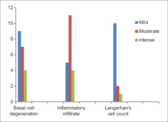
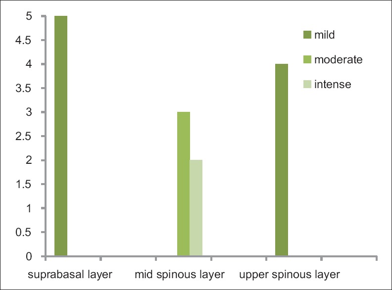
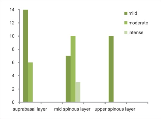
The average percentage of CD1a-positive LCs in normal subjects was 0.9%, and in the OLP cases higher percentage was observed (3.93%). The statistical comparison of these two parameters was significant (=0.018). The degree of basal cell degeneration and density of subepithelial infiltrate on statistical comparison with the concentration of CD1a-positive LCs showed significant results.

CONCLUSION

LCs play a pivotal role in the recruitment of CD4+ and CD8+ cells to the subepithelial region and basal keratinocytes apoptosis. A small number of study subjects, assessment of only CD1a molecule and LCs in the epidermis only were a few of the drawbacks of the study.

摘要

背景/目的:扁平苔藓是一种T细胞介导的黏膜皮肤疾病,其组织病理学特征为上皮下区有一条慢性炎症细胞带以及基底层变性。本研究旨在评估口腔扁平苔藓(OLP)中分化簇1a(CD1a)阳性朗格汉斯细胞(LCs)的分布及定量评估,从而确定LCs在OLP发生变化中的作用。

材料与方法

用CD1a抗体对5例正常口腔黏膜和20例OLP进行免疫染色;在显微照片中手动计数阳性细胞,并使用t检验、曼-惠特尼检验和威尔科克森符号秩检验进行统计分析。

结果

正常受试者中CD1a阳性LCs的平均百分比为0.9%,而在OLP病例中观察到更高的百分比(3.93%)。这两个参数的统计比较具有显著性(P=0.018)。与CD1a阳性LCs浓度进行统计比较时,基底细胞变性程度和上皮下浸润密度显示出显著结果。

结论

LCs在CD4+和CD8+细胞募集到上皮下区域以及基底角质形成细胞凋亡中起关键作用。研究对象数量少、仅评估表皮中的CD1a分子和LCs是该研究的一些不足之处。

https://cdn.ncbi.nlm.nih.gov/pmc/blobs/3579/6340233/7a10ff02cd34/IJD-64-41-g006.jpg
https://cdn.ncbi.nlm.nih.gov/pmc/blobs/3579/6340233/6929bc5af42e/IJD-64-41-g001.jpg
https://cdn.ncbi.nlm.nih.gov/pmc/blobs/3579/6340233/25414bc407f1/IJD-64-41-g002.jpg
https://cdn.ncbi.nlm.nih.gov/pmc/blobs/3579/6340233/1695e427bd60/IJD-64-41-g003.jpg
https://cdn.ncbi.nlm.nih.gov/pmc/blobs/3579/6340233/d75bb265d77f/IJD-64-41-g004.jpg
https://cdn.ncbi.nlm.nih.gov/pmc/blobs/3579/6340233/23a2357f7156/IJD-64-41-g005.jpg
https://cdn.ncbi.nlm.nih.gov/pmc/blobs/3579/6340233/7a10ff02cd34/IJD-64-41-g006.jpg

相似文献

1
Expression of Cluster of Differentiation 1a-Positive Langerhans Cells in Oral Lichen Planus.口腔扁平苔藓中分化簇1a阳性朗格汉斯细胞的表达
Indian J Dermatol. 2019 Jan-Feb;64(1):41-46. doi: 10.4103/ijd.IJD_350_16.
2
Expression of CD1a by Langerhan's Cells in Oral Lichen Planus - A Retrospective Analysis.口腔扁平苔藓中朗格汉斯细胞CD1a的表达——一项回顾性分析
J Clin Diagn Res. 2016 Jun;10(6):ZC28-31. doi: 10.7860/JCDR/2016/19189.7966. Epub 2016 Jun 1.
3
Immunohistochemical evaluation of Langerhans cells in oral lichen planus and oral lichenoid lesions.口腔扁平苔藓和口腔类扁平苔藓病变中朗格汉斯细胞的免疫组织化学评估。
Arch Oral Biol. 2021 Apr;124:105027. doi: 10.1016/j.archoralbio.2020.105027. Epub 2020 Dec 8.
4
A Comparative Immunohistochemical Analysis of Langerhans Cells in Oral Mucosa, Oral Lichen Planus and Oral Squamous Cell Carcinoma.口腔黏膜、口腔扁平苔藓和口腔鳞状细胞癌中朗格汉斯细胞的比较免疫组织化学分析
J Clin Diagn Res. 2015 Jul;9(7):ZC76-9. doi: 10.7860/JCDR/2015/14170.6235. Epub 2015 Jul 1.
5
Langerin-expressing and CD83-expressing cells in oral lichen planus lesions.口腔扁平苔藓病变中表达朗格汉斯蛋白和表达CD83的细胞。
Acta Odontol Scand. 2007 Jun;65(3):156-61. doi: 10.1080/00016350601137251.
6
Oral lichen planus: immunohistology of mucosal lesions.口腔扁平苔藓:黏膜病变的免疫组织学
J Oral Pathol Med. 2002 Aug;31(7):410-4. doi: 10.1034/j.1600-0714.2002.00097.x.
7
Langerhans Cells, T Cells, and B Cells in Oral Lichen Planus and Oral Leukoplakia.口腔扁平苔藓和口腔白斑中的朗格汉斯细胞、T细胞和B细胞。
Int J Dent. 2022 Mar 22;2022:5430309. doi: 10.1155/2022/5430309. eCollection 2022.
8
Langerhans cells and T cells in oral graft versus host disease and oral lichen planus.口腔移植物抗宿主病和口腔扁平苔藓中的朗格汉斯细胞和T细胞。
Scand J Immunol. 2001 Nov;54(5):516-24. doi: 10.1046/j.1365-3083.2001.00988.x.
9
Immunohistochemical Study of p53 Expression in Patients with Erosive and Non-Erosive Oral Lichen Planus.糜烂性和非糜烂性口腔扁平苔藓患者p53表达的免疫组织化学研究
J Dent (Shiraz). 2018 Jun;19(2):118-123.
10
Apoptosis in oral lichen planus.口腔扁平苔藓中的细胞凋亡
Eur J Oral Sci. 2001 Oct;109(5):361-4. doi: 10.1034/j.1600-0722.2001.00081.x.

引用本文的文献

1
CD8 + T-cell marker genes reveal different immune subtypes of oral lichen planus by integrating single-cell RNA-seq and bulk RNA-sequencing.CD8+T 细胞标记基因通过整合单细胞 RNA-seq 和批量 RNA-seq 揭示口腔扁平苔藓的不同免疫亚型。
BMC Oral Health. 2023 Jul 8;23(1):464. doi: 10.1186/s12903-023-03138-0.
2
Updates on immunological mechanistic insights and targeting of the oral lichen planus microenvironment.口腔扁平苔藓微环境的免疫机制见解和靶向治疗的最新进展。
Front Immunol. 2023 Jan 9;13:1023213. doi: 10.3389/fimmu.2022.1023213. eCollection 2022.
3
FoxP3CD4, IFN-γCD4, and IFN-γCD8 cell levels in erosive and non-erosive types of oral lichen planus patients.

本文引用的文献

1
Pathophysiology of Langerhans cells.朗格汉斯细胞的病理生理学
J Oral Maxillofac Pathol. 2012 May;16(2):239-44. doi: 10.4103/0973-029X.99077.
2
Increased number of Langerhans cells in oral lichen planus and oral lichenoid lesions.口腔扁平苔藓和口腔类扁平苔藓病中朗格汉斯细胞数量增加。
Oral Surg Oral Med Oral Pathol Oral Radiol. 2012 May;113(5):661-6. doi: 10.1016/j.oooo.2011.12.008. Epub 2012 Apr 12.
3
Pathophysiology, etiologic factors, and clinical management of oral lichen planus, part I: facts and controversies.口腔扁平苔藓的病理生理学、病因因素和临床管理,第一部分:事实和争议。
糜烂型和非糜烂型口腔扁平苔藓患者的FoxP3 CD4、IFN-γ CD4和IFN-γ CD8细胞水平。
J Dent Sci. 2021 Mar;16(2):751-756. doi: 10.1016/j.jds.2021.01.005. Epub 2021 Jan 27.
4
Marking vertebrates langerhans cells, from fish to mammals.标记鱼类到哺乳动物的朗格汉斯细胞。
Acta Histochem. 2020 Oct;122(7):151622. doi: 10.1016/j.acthis.2020.151622. Epub 2020 Sep 9.
Clin Dermatol. 2010 Jan-Feb;28(1):100-8. doi: 10.1016/j.clindermatol.2009.03.004.
4
Lichenoid tissue reaction/interface dermatitis: clinical and histological perspectives.苔藓样组织反应/界面性皮炎:临床与组织学视角
J Invest Dermatol. 2009 May;129(5):1088-99. doi: 10.1038/sj.jid.2009.42. Epub 2009 Feb 26.
5
Interstitial and Langerhans' dendritic cells in chronic periodontitis and gingivitis.慢性牙周炎和牙龈炎中的间质细胞和朗格汉斯树突状细胞。
Braz Oral Res. 2008 Jul-Sep;22(3):258-63. doi: 10.1590/s1806-83242008000300012.
6
Activin A induces Langerhans cell differentiation in vitro and in human skin explants.激活素A在体外和人皮肤外植体中诱导朗格汉斯细胞分化。
PLoS One. 2008 Sep 24;3(9):e3271. doi: 10.1371/journal.pone.0003271.
7
Oral lichen planus: clinical and histopathological considerations.口腔扁平苔藓:临床与组织病理学考量
Braz J Otorhinolaryngol. 2008 Mar-Apr;74(2):284-92. doi: 10.1016/s1808-8694(15)31102-2.
8
Oral lichen planus and lichenoid reactions: etiopathogenesis, diagnosis, management and malignant transformation.口腔扁平苔藓和苔藓样反应:病因发病机制、诊断、治疗及恶变
J Oral Sci. 2007 Jun;49(2):89-106. doi: 10.2334/josnusd.49.89.
9
Langerin is a natural barrier to HIV-1 transmission by Langerhans cells.朗格汉斯细胞型凝集素是朗格汉斯细胞对HIV-1传播的天然屏障。
Nat Med. 2007 Mar;13(3):367-71. doi: 10.1038/nm1541. Epub 2007 Mar 4.
10
Psychological profile in oral lichen planus.口腔扁平苔藓的心理特征
J Clin Periodontol. 2005 Oct;32(10):1034-40. doi: 10.1111/j.1600-051X.2005.00829.x.