Li Yuefeng, Zhou Xiaofen, Zhang Qiongying, Chen Endong, Sun Yihan, Ye Danrong, Wang Ouchen, Zhang Xiaohua, Lyu Jianxin
Key Laboratory of Laboratory Medicine, Ministry of Education, Zhejiang Provincial Key Laboratory of Medical Genetics, College of Laboratory Medicine and Life Sciences, Wenzhou Medical University, Wenzhou, Zhejiang, China,
Department of Thyroid and Breast Surgery, The First Affiliated Hospital of Wenzhou Medical University, Wenzhou, Zhejiang, China,
Cancer Manag Res. 2019 Jan 22;11:931-941. doi: 10.2147/CMAR.S183355. eCollection 2019.
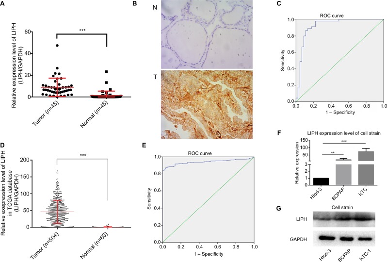
Papillary thyroid carcinoma (PTC) is the most common type of thyroid carcinoma, which is associated with a high incidence of lymph-node metastasis. Multiple biomarkers have been identified for the precise diagnosis of PTC at an early stage. However, their role in PTC remains poorly elucidated. Previously, we reported that lipase H (LIPH), a membrane-bound protein, was highly expressed in PTC. This study aimed to fully elucidate the causal role of in the development of PTC and investigated its relationship with lymph-node metastasis in PTC.
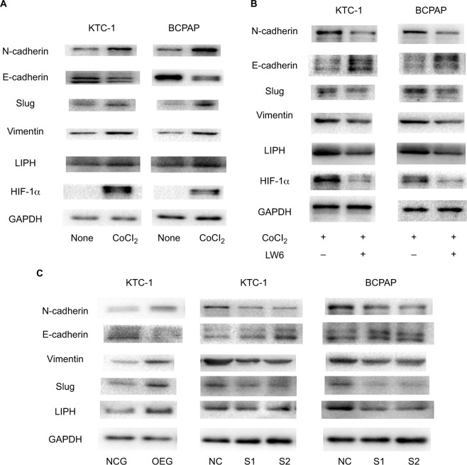
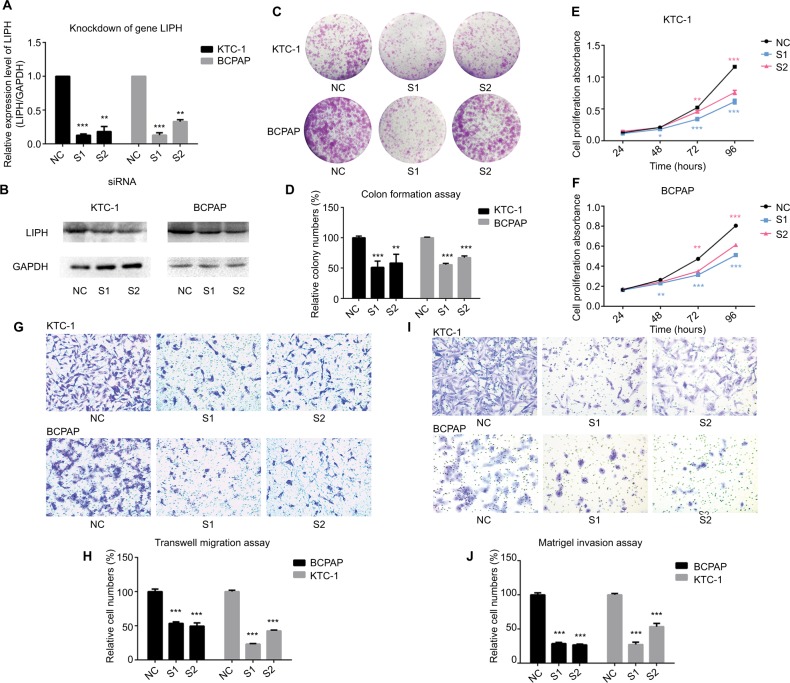
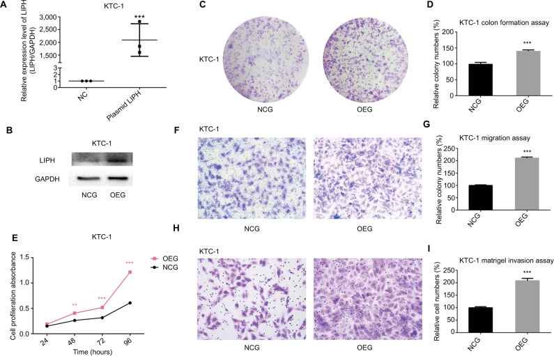
Quantitative reverse transcription PCR and immunohistochemistry were used to measure the mRNA and protein expression levels of in 45 and 6 pairs of PTC tissues and adjacent normal tissues, respectively. Clinical tissue data of 504 PTC tissues and 60 normal thyroid tissues from The Cancer Genome Atlas database were used to analyze the correlation between expression level and clinical features in PTC. siRNAs were used to knock down genes, while plasmids were used to overexpress genes. Two PTC cell lines (KTC-1 and BCPAP) were used in subsequent cytological function studies. In addition, a hypoxia stress model was constructed using cobaltous chloride hexahydrate reagent, and the protein expression level of the corresponding biomarkers was measured by Western blotting.
This study revealed that high expression of in PTC was closely associated with lymph-node metastasis. Our cellular function experiments indicated that positively correlated with the malignant behavior of PTC cell lines. We further confirmed the role of in hypoxia and its relationship with the epithelial-mesenchymal transition pathway in PTC.
plays an important role in PTC oncogenesis and development, especially in lymph-node metastasis. It can be regarded as a biomarker for the diagnosis and treatment of PTC in the near future.
甲状腺乳头状癌(PTC)是最常见的甲状腺癌类型,与高淋巴结转移发生率相关。已鉴定出多种生物标志物用于PTC的早期精确诊断。然而,它们在PTC中的作用仍未得到充分阐明。此前,我们报道脂肪酶H(LIPH),一种膜结合蛋白,在PTC中高表达。本研究旨在全面阐明其在PTC发生发展中的因果作用,并研究其与PTC淋巴结转移的关系。
分别采用定量逆转录PCR和免疫组织化学方法检测45对和6对PTC组织及相邻正常组织中该蛋白的mRNA和蛋白表达水平。利用来自癌症基因组图谱数据库的504例PTC组织和60例正常甲状腺组织的临床组织数据,分析该蛋白表达水平与PTC临床特征之间的相关性。使用小干扰RNA(siRNAs)敲低基因,同时使用质粒过表达基因。随后的细胞学功能研究使用了两种PTC细胞系(KTC-1和BCPAP)。此外,使用六水合氯化钴试剂构建缺氧应激模型,并通过蛋白质印迹法检测相应生物标志物的蛋白表达水平。
本研究表明,PTC中该蛋白的高表达与淋巴结转移密切相关。我们的细胞功能实验表明,该蛋白与PTC细胞系的恶性行为呈正相关。我们进一步证实了该蛋白在缺氧中的作用及其与PTC上皮-间质转化途径的关系。
该蛋白在PTC的发生发展中起重要作用,尤其是在淋巴结转移方面。在不久的将来,它可被视为PTC诊断和治疗中的一种生物标志物。