• 文献检索
  • 文档翻译
  • 深度研究
  • 学术资讯
  • Suppr Zotero 插件Zotero 插件
  • 邀请有礼
  • 套餐&价格
  • 历史记录
应用&插件
Suppr Zotero 插件Zotero 插件浏览器插件Mac 客户端Windows 客户端微信小程序
定价
高级版会员购买积分包购买API积分包
服务
文献检索文档翻译深度研究API 文档MCP 服务
关于我们
关于 Suppr公司介绍联系我们用户协议隐私条款
关注我们

Suppr 超能文献

核心技术专利:CN118964589B侵权必究
粤ICP备2023148730 号-1Suppr @ 2026

文献检索

告别复杂PubMed语法,用中文像聊天一样搜索,搜遍4000万医学文献。AI智能推荐,让科研检索更轻松。

立即免费搜索

文件翻译

保留排版,准确专业,支持PDF/Word/PPT等文件格式,支持 12+语言互译。

免费翻译文档

深度研究

AI帮你快速写综述,25分钟生成高质量综述,智能提取关键信息,辅助科研写作。

立即免费体验

急性和慢性单侧及双侧丘脑病变的神经影像学

Neuroimaging of acute and chronic unilateral and bilateral thalamic lesions.

作者信息

Tuttle C, Boto J, Martin S, Barnaure I, Korchi A M, Scheffler M, Vargas M I

机构信息

Division of Radiology, Faculty of Medicine, Geneva University Hospital, Geneva, Switzerland.

Division of Neuroradiology, DISIM, Faculty of Medicine, Geneva University Hospital, Rue Gabrielle-Perret-Gentil 4, 1211, Geneva 14, Switzerland.

出版信息

Insights Imaging. 2019 Feb 22;10(1):24. doi: 10.1186/s13244-019-0700-3.

DOI:10.1186/s13244-019-0700-3
PMID:30796553
原文链接:https://pmc.ncbi.nlm.nih.gov/articles/PMC6386756/
Abstract

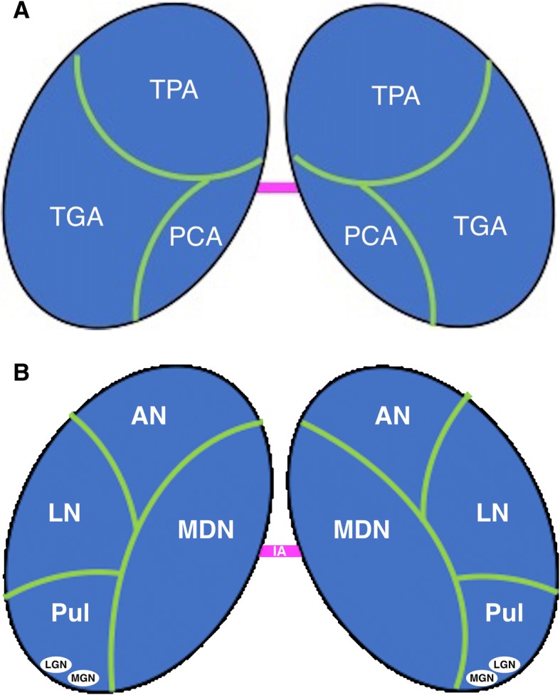
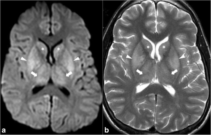
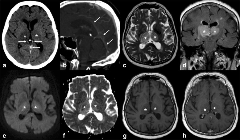
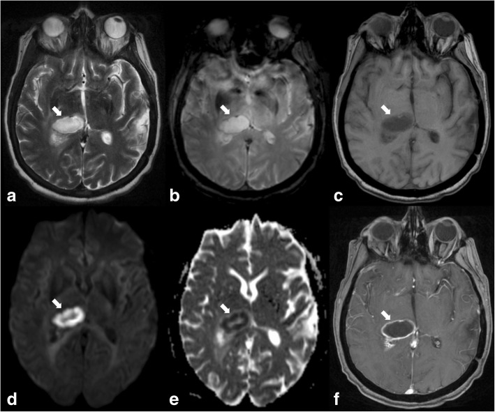
The thalami are bilateral ovoid grey matter cerebral structures bordering the third ventricle on both sides, which participate in functions such as relaying of sensory and motor signals, regulation of consciousness, and alertness. Pathologies affecting the thalami can be of neoplastic, infectious, vascular, toxic, metabolic, or congenital origin.The purpose of this review is to provide a comprehensive approach to the thalamus focusing on its anatomy, the main pathologies affecting this structure and their radiological semiology on CT and MRI. We will also illustrate the importance of multimodal MR imaging (morphologic sequences, diffusion-weighted imaging, perfusion, spectroscopy) for the diagnosis and treatment of these conditions.

摘要

丘脑是双侧的卵圆形灰质脑结构,两侧与第三脑室相邻,参与感觉和运动信号的传递、意识调节及警觉等功能。影响丘脑的病变可能源于肿瘤、感染、血管、中毒、代谢或先天性因素。本综述的目的是提供一种全面探讨丘脑的方法,重点关注其解剖结构、影响该结构的主要病变及其在CT和MRI上的放射学征象。我们还将阐述多模态磁共振成像(形态学序列、扩散加权成像、灌注成像、波谱分析)在这些疾病诊断和治疗中的重要性。

https://cdn.ncbi.nlm.nih.gov/pmc/blobs/e1f6/6386756/e72ac3ec1abd/13244_2019_700_Fig15_HTML.jpg
https://cdn.ncbi.nlm.nih.gov/pmc/blobs/e1f6/6386756/9d34e9fba966/13244_2019_700_Fig1_HTML.jpg
https://cdn.ncbi.nlm.nih.gov/pmc/blobs/e1f6/6386756/0684594bdfbc/13244_2019_700_Fig2_HTML.jpg
https://cdn.ncbi.nlm.nih.gov/pmc/blobs/e1f6/6386756/2eb238198449/13244_2019_700_Fig3_HTML.jpg
https://cdn.ncbi.nlm.nih.gov/pmc/blobs/e1f6/6386756/922ad22d6857/13244_2019_700_Fig4_HTML.jpg
https://cdn.ncbi.nlm.nih.gov/pmc/blobs/e1f6/6386756/5ce0567757c7/13244_2019_700_Fig5_HTML.jpg
https://cdn.ncbi.nlm.nih.gov/pmc/blobs/e1f6/6386756/71220336f392/13244_2019_700_Fig6_HTML.jpg
https://cdn.ncbi.nlm.nih.gov/pmc/blobs/e1f6/6386756/526455785e74/13244_2019_700_Fig7_HTML.jpg
https://cdn.ncbi.nlm.nih.gov/pmc/blobs/e1f6/6386756/d93f8e83153f/13244_2019_700_Fig8_HTML.jpg
https://cdn.ncbi.nlm.nih.gov/pmc/blobs/e1f6/6386756/dd152881b129/13244_2019_700_Fig9_HTML.jpg
https://cdn.ncbi.nlm.nih.gov/pmc/blobs/e1f6/6386756/158ecbbf124f/13244_2019_700_Fig10_HTML.jpg
https://cdn.ncbi.nlm.nih.gov/pmc/blobs/e1f6/6386756/a5321893a95c/13244_2019_700_Fig11_HTML.jpg
https://cdn.ncbi.nlm.nih.gov/pmc/blobs/e1f6/6386756/f98ed83f51f1/13244_2019_700_Fig12_HTML.jpg
https://cdn.ncbi.nlm.nih.gov/pmc/blobs/e1f6/6386756/da208bef00dc/13244_2019_700_Fig13_HTML.jpg
https://cdn.ncbi.nlm.nih.gov/pmc/blobs/e1f6/6386756/cea29adee793/13244_2019_700_Fig14_HTML.jpg
https://cdn.ncbi.nlm.nih.gov/pmc/blobs/e1f6/6386756/e72ac3ec1abd/13244_2019_700_Fig15_HTML.jpg

相似文献

1
Neuroimaging of acute and chronic unilateral and bilateral thalamic lesions.急性和慢性单侧及双侧丘脑病变的神经影像学
Insights Imaging. 2019 Feb 22;10(1):24. doi: 10.1186/s13244-019-0700-3.
2
Bilateral lesions of the basal ganglia and thalami (central grey matter)-pictorial review.双侧基底节和丘脑病变(中央灰质)——图像综述。
Neuroradiology. 2020 Dec;62(12):1565-1605. doi: 10.1007/s00234-020-02511-y. Epub 2020 Aug 5.
3
Bilateral thalamic lesions: A pictorial review.双侧丘脑病变:影像综述
J Med Imaging Radiat Oncol. 2017 Jun;61(3):353-360. doi: 10.1111/1754-9485.12597. Epub 2017 Feb 23.
4
The value of morphological neuroimaging after acute exposure to toxic substances.急性接触有毒物质后形态学神经影像学的价值。
Toxicol Rev. 2006;25(2):87-98. doi: 10.2165/00139709-200625020-00003.
5
Diffusion-weighted MR of the brain: methodology and clinical application.脑部扩散加权磁共振成像:方法与临床应用
Radiol Med. 2005 Mar;109(3):155-97.
6
[Early assessment of severe hypoxic-ischemic encephalopathy in neonates by diffusion-weighted magnetic resonance imaging techniques and its significance].[磁共振扩散加权成像技术对新生儿重度缺氧缺血性脑病的早期评估及其意义]
Zhonghua Er Ke Za Zhi. 2007 Nov;45(11):843-7.
7
Imaging bithalamic pathology in the pediatric brain: demystifying a diagnostic conundrum.双侧丘脑病变的儿科脑影像学:解开诊断难题。
AJR Am J Roentgenol. 2011 Dec;197(6):1449-59. doi: 10.2214/AJR.11.6702.
8
Bilateral thalamic tumors in children.儿童双侧丘脑肿瘤。
Childs Nerv Syst. 2002 Aug;18(8):440-4. doi: 10.1007/s00381-002-0609-9. Epub 2002 Jul 2.
9
Midbrain and bilateral paramedian thalamic stroke due to artery of Percheron occlusion.由于Percheron动脉闭塞导致的中脑和双侧丘脑旁正中梗死。
Neurol Neurochir Pol. 2016;50(3):180-4. doi: 10.1016/j.pjnns.2016.01.008. Epub 2016 Jan 24.
10
Neuropathology of Mild Traumatic Brain Injury: Correlation to Neurocognitive and Neurobehavioral Findings轻度创伤性脑损伤的神经病理学:与神经认知和神经行为结果的相关性

引用本文的文献

1
Endovascular Management of Arteriovenous Malformations in the Thalamic and Basal Ganglia: A Systematic Review.丘脑和基底节区动静脉畸形的血管内治疗:一项系统评价
Cureus. 2025 Jan 5;17(1):e76933. doi: 10.7759/cureus.76933. eCollection 2025 Jan.
2
Incidental Thalamic Lesions Identified on Brain MRI in Pediatric and Young Adult Patients: Imaging Features and Natural History.脑 MRI 检查中偶然发现的儿童和青年患者的丘脑病变:影像学特征和自然病史。
AJNR Am J Neuroradiol. 2024 Feb 7;45(2):211-217. doi: 10.3174/ajnr.A8090.
3
Dural Arteriovenous Fistula Presenting as a Rapidly Progressive Thalamic Dementia: A Case Report.

本文引用的文献

1
Clinical applications of diffusion weighted imaging in neuroradiology.扩散加权成像在神经放射学中的临床应用。
Insights Imaging. 2018 Aug;9(4):535-547. doi: 10.1007/s13244-018-0624-3. Epub 2018 May 30.
2
Localization of Deep Brain Stimulation Contacts Using Corticospinal/Corticobulbar Tracts Stimulation.利用皮质脊髓/皮质延髓束刺激对脑深部电刺激触点进行定位
Front Neurol. 2017 May 31;8:239. doi: 10.3389/fneur.2017.00239. eCollection 2017.
3
Evaluation of electrode position in deep brain stimulation by image fusion (MRI and CT).
表现为快速进展性丘脑痴呆的硬脑膜动静脉瘘:一例报告
Cureus. 2022 Sep 21;14(9):e29392. doi: 10.7759/cureus.29392. eCollection 2022 Sep.
4
Acute percheron infarction: a precision learning.急性佩尔戎梗死:精准学习。
BMC Neurol. 2022 Jun 4;22(1):207. doi: 10.1186/s12883-022-02735-w.
5
Bilateral Thalamic Glioma: A Case Report.双侧丘脑胶质瘤:一例报告
Cureus. 2021 Nov 14;13(11):e19570. doi: 10.7759/cureus.19570. eCollection 2021 Nov.
6
Bilateral thalamic changes in anti-NMDAR encephalitis presenting with hemichorea and dystonia and acute transient psychotic disorder.抗 NMDAR 脑炎表现为偏侧舞蹈症和肌张力障碍伴急性短暂性精神病性障碍的双侧丘脑改变。
J Neuroimmunol. 2020 Oct 15;347:577329. doi: 10.1016/j.jneuroim.2020.577329. Epub 2020 Jul 22.
7
Bilateral thalamic lesions in a patient with probable acute disseminating encephalomyelitis: a case report.双侧丘脑病变一例疑似急性播散性脑脊髓炎患者:病例报告。
BMC Neurol. 2020 Jul 1;20(1):265. doi: 10.1186/s12883-020-01834-w.
8
The Perirolandic Sign: A Unique Imaging Finding Observed in Association with Polymerase γ-Related Disorders.类岛带征:一种与聚合酶γ相关疾病相关的独特影像学发现。
AJNR Am J Neuroradiol. 2020 May;41(5):917-922. doi: 10.3174/ajnr.A6514. Epub 2020 May 7.
通过图像融合(MRI和CT)评估深部脑刺激中的电极位置。
Neuroradiology. 2015 Sep;57(9):903-8. doi: 10.1007/s00234-015-1547-z. Epub 2015 May 29.
4
Anterior thalamic nuclei lesions and recovery of function: Relevance to cognitive thalamus.丘脑前核病变与功能恢复:与认知丘脑的相关性
Neurosci Biobehav Rev. 2015 Jul;54:145-60. doi: 10.1016/j.neubiorev.2014.12.007. Epub 2015 Jan 29.
5
The fornix and limbic system.穹窿和边缘系统。
Semin Ultrasound CT MR. 2014 Oct;35(5):459-73. doi: 10.1053/j.sult.2014.06.005. Epub 2014 Jun 25.
6
Illustrating the relevance of updated diagnostic criteria for sporadic Creutzfeldt-Jakob disease: a teaching neurocase.说明散发性克雅氏病更新诊断标准的相关性:一个教学神经病例。
BMJ Case Rep. 2013 Aug 28;2013:bcr2013010061. doi: 10.1136/bcr-2013-010061.
7
Percentage signal recovery derived from MR dynamic susceptibility contrast imaging is useful to differentiate common enhancing malignant lesions of the brain.基于磁共振动态磁敏感对比成像的信号恢复百分比有助于鉴别常见的脑实质增强性恶性病变。
AJNR Am J Neuroradiol. 2011 Jun-Jul;32(6):1004-10. doi: 10.3174/ajnr.A2441. Epub 2011 Apr 21.
8
Artery of percheron infarction: imaging patterns and clinical spectrum.Percheron 梗死动脉:影像学表现及临床谱。
AJNR Am J Neuroradiol. 2010 Aug;31(7):1283-9. doi: 10.3174/ajnr.A2044. Epub 2010 Mar 18.
9
Bilateral thalamic lesions.双侧丘脑病变。
AJR Am J Roentgenol. 2009 Feb;192(2):W53-62. doi: 10.2214/AJR.08.1585.
10
Advanced MR imaging techniques in the diagnosis of intraaxial brain tumors in adults.成人脑内肿瘤诊断中的先进磁共振成像技术
Radiographics. 2006 Oct;26 Suppl 1:S173-89. doi: 10.1148/rg.26si065513.