Madani Ali, Arnaout Ramy, Mofrad Mohammad, Arnaout Rima
Molecular Biophysics and Integrated Bioimaging Division, Lawrence Berkeley National Lab, California Institute for Quantitative Biosciences (QB3), University of California at Berkeley, 208A Stanley Hall Room 1762, Berkeley, CA 94720, USA.
Beth Israel Deaconess Medical Center, Harvard Medical School, 330 Brookline Avenue Dana 615, Boston, MA 02215, USA.
NPJ Digit Med. 2018;1. doi: 10.1038/s41746-017-0013-1. Epub 2018 Mar 21.
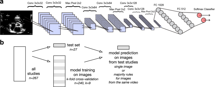
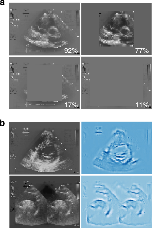
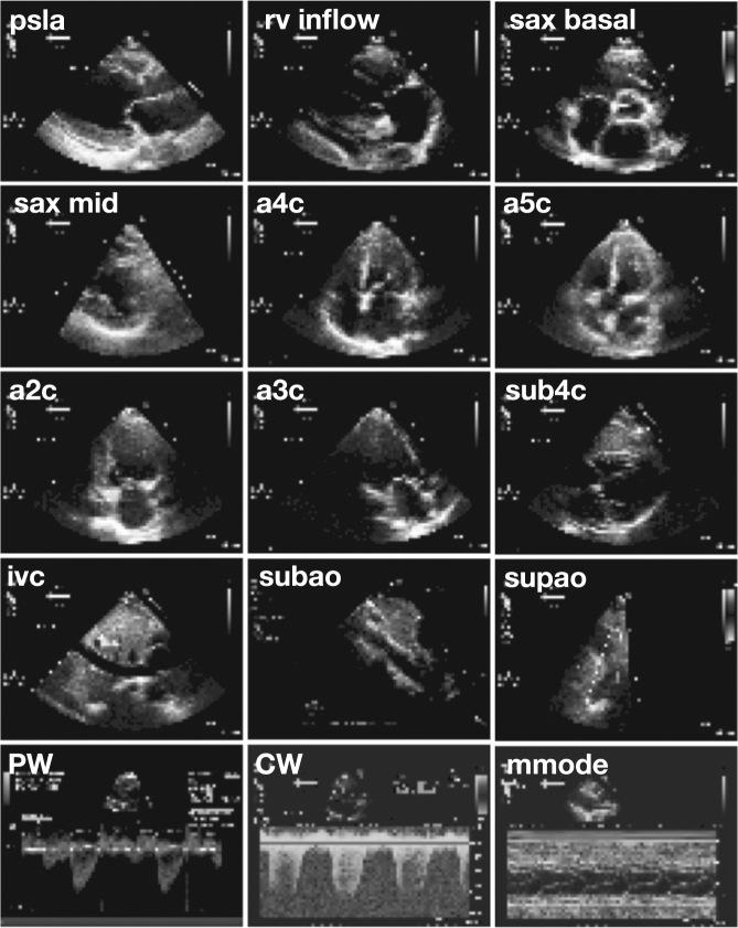
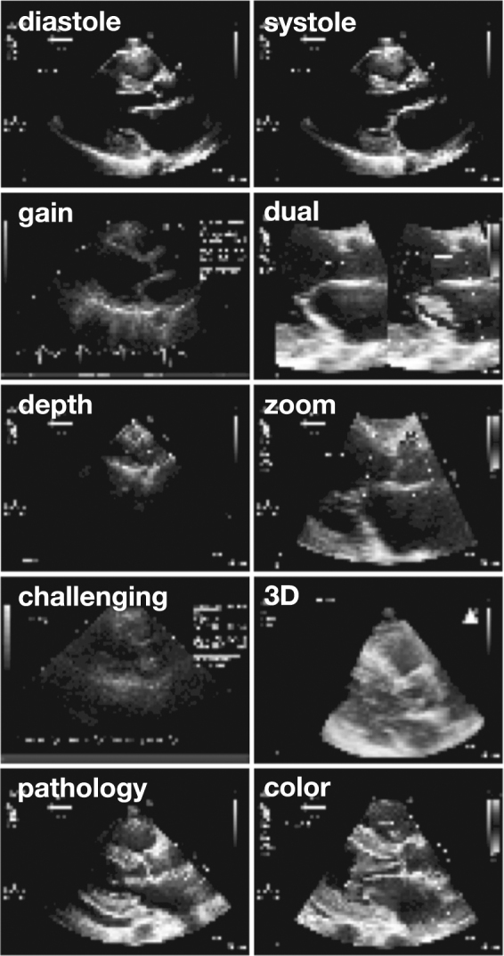
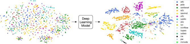
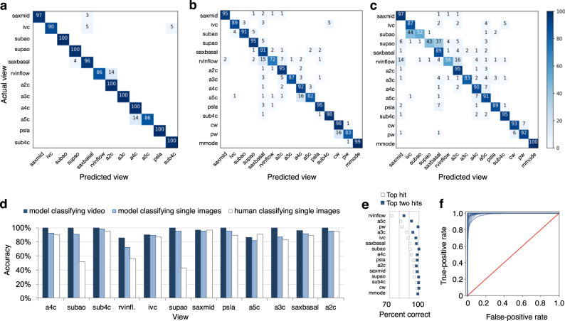
Echocardiography is essential to cardiology. However, the need for human interpretation has limited echocardiography's full potential for precision medicine. Deep learning is an emerging tool for analyzing images but has not yet been widely applied to echocardiograms, partly due to their complex multi-view format. The essential first step toward comprehensive computer-assisted echocardiographic interpretation is determining whether computers can learn to recognize these views. We trained a convolutional neural network to simultaneously classify 15 standard views (12 video, 3 still), based on labeled still images and videos from 267 transthoracic echocardiograms that captured a range of real-world clinical variation. Our model classified among 12 video views with 97.8% overall test accuracy without overfitting. Even on single low-resolution images, accuracy among 15 views was 91.7% vs. 70.2-84.0% for board-certified echocardiographers. Data visualization experiments showed that the model recognizes similarities among related views and classifies using clinically relevant image features. Our results provide a foundation for artificial intelligence-assisted echocardiographic interpretation.
超声心动图对心脏病学至关重要。然而,由于需要人工解读,超声心动图在精准医疗方面的全部潜力受到了限制。深度学习是一种新兴的图像分析工具,但尚未广泛应用于超声心动图,部分原因是其复杂的多视图格式。迈向全面计算机辅助超声心动图解读的关键第一步是确定计算机是否能够学会识别这些视图。我们基于来自267份经胸超声心动图的标记静态图像和视频训练了一个卷积神经网络,这些图像和视频捕捉了一系列真实世界的临床变化,用于同时对15个标准视图(12个视频视图、3个静态视图)进行分类。我们的模型在12个视频视图中的总体测试准确率为97.8%,且没有过拟合。即使是在单个低分辨率图像上,15个视图的准确率为91.7%,而经委员会认证的超声心动图专家的准确率为70.2 - 84.0%。数据可视化实验表明,该模型能够识别相关视图之间的相似性,并利用临床相关的图像特征进行分类。我们的结果为人工智能辅助超声心动图解读奠定了基础。