• 文献检索
  • 文档翻译
  • 深度研究
  • 学术资讯
  • Suppr Zotero 插件Zotero 插件
  • 邀请有礼
  • 套餐&价格
  • 历史记录
应用&插件
Suppr Zotero 插件Zotero 插件浏览器插件Mac 客户端Windows 客户端微信小程序
定价
高级版会员购买积分包购买API积分包
服务
文献检索文档翻译深度研究API 文档MCP 服务
关于我们
关于 Suppr公司介绍联系我们用户协议隐私条款
关注我们

Suppr 超能文献

核心技术专利:CN118964589B侵权必究
粤ICP备2023148730 号-1Suppr @ 2026

文献检索

告别复杂PubMed语法,用中文像聊天一样搜索,搜遍4000万医学文献。AI智能推荐,让科研检索更轻松。

立即免费搜索

文件翻译

保留排版,准确专业,支持PDF/Word/PPT等文件格式,支持 12+语言互译。

免费翻译文档

深度研究

AI帮你快速写综述,25分钟生成高质量综述,智能提取关键信息,辅助科研写作。

立即免费体验

羧基化量子点在人骨髓间充质干细胞中的摄取和分布:细胞生长密度很重要。

Uptake and distribution of carboxylated quantum dots in human mesenchymal stem cells: cell growing density matters.

机构信息

Biobank, National Cancer Institute, Baublio Str. 3b, 08406, Vilnius, Lithuania.

Laboratory of Immunology, National Cancer Institute, Baublio Str. 3b, 08406, Vilnius, Lithuania.

出版信息

J Nanobiotechnology. 2019 Mar 13;17(1):39. doi: 10.1186/s12951-019-0470-6.

DOI:10.1186/s12951-019-0470-6
PMID:30866960
原文链接:https://pmc.ncbi.nlm.nih.gov/articles/PMC6417192/
Abstract

BACKGROUND

Human mesenchymal stem cells (MSCs) have drawn much attention in the field of regenerative medicine for their immunomodulatory and anti-inflammatory effects. MSCs possess specific tumor-oriented migration and incorporation highlighting the potential for MSCs to be used as an ideal carrier for anticancer agents. Bone marrow is the main source of MSCs for clinical applications. MSCs tracking in vivo is a critical component of the safety and efficacy evaluation of therapeutic cell products; therefore, cells must be labeled with contrast agents to enable visualization of the MSCs migration in vivo. Due to their unique properties, quantum dots (QDs) are emerging as optimal tools in long-term MSC optical imaging applications. The aim of this study was to investigate the uptake dynamics, cytotoxity, subcellular and extracellular distribution of non-targeted carboxylated quantum dots in human bone marrow MSCs at different cell growing densities.

RESULTS

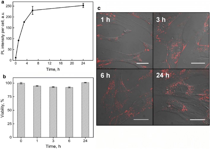
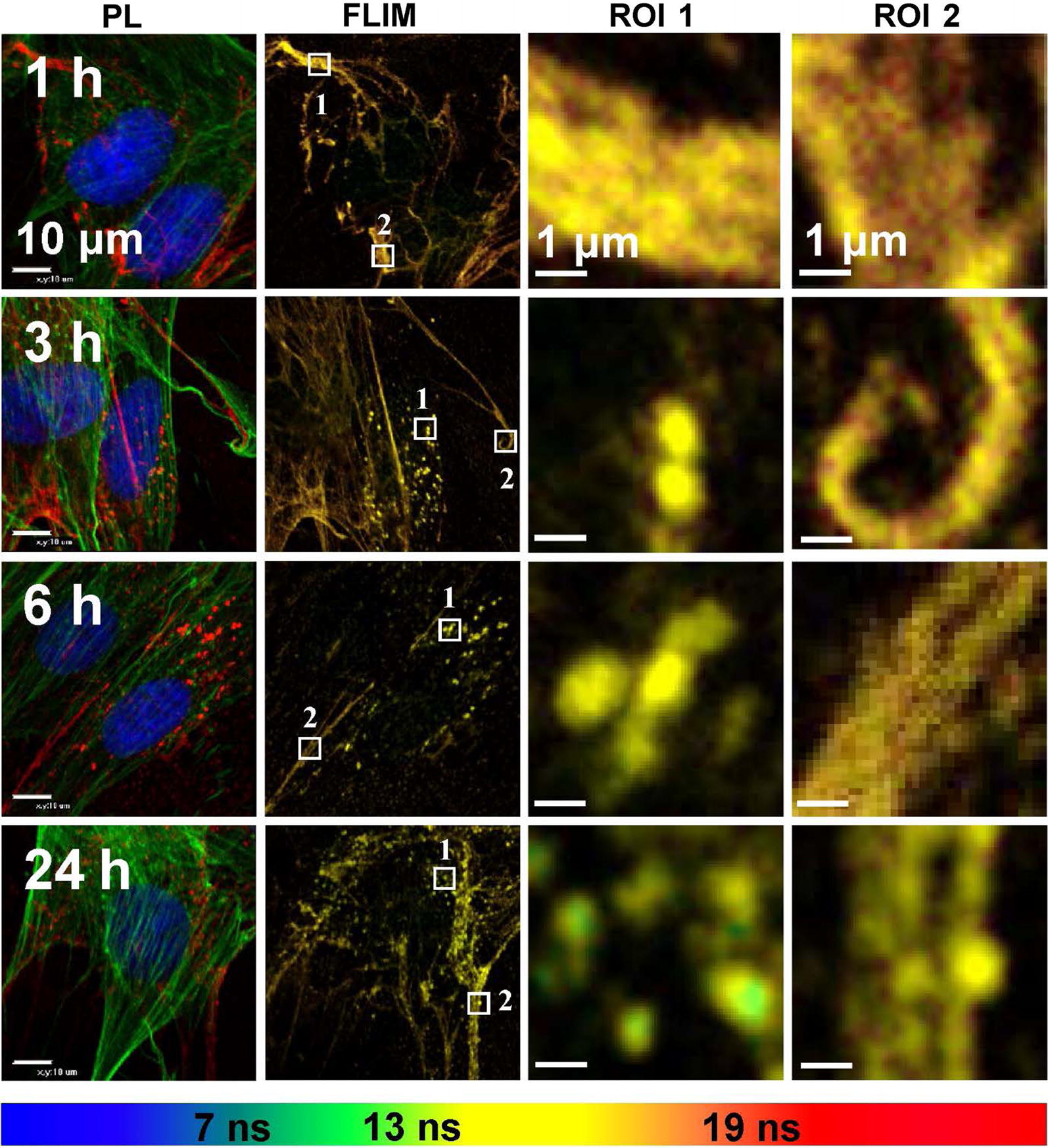
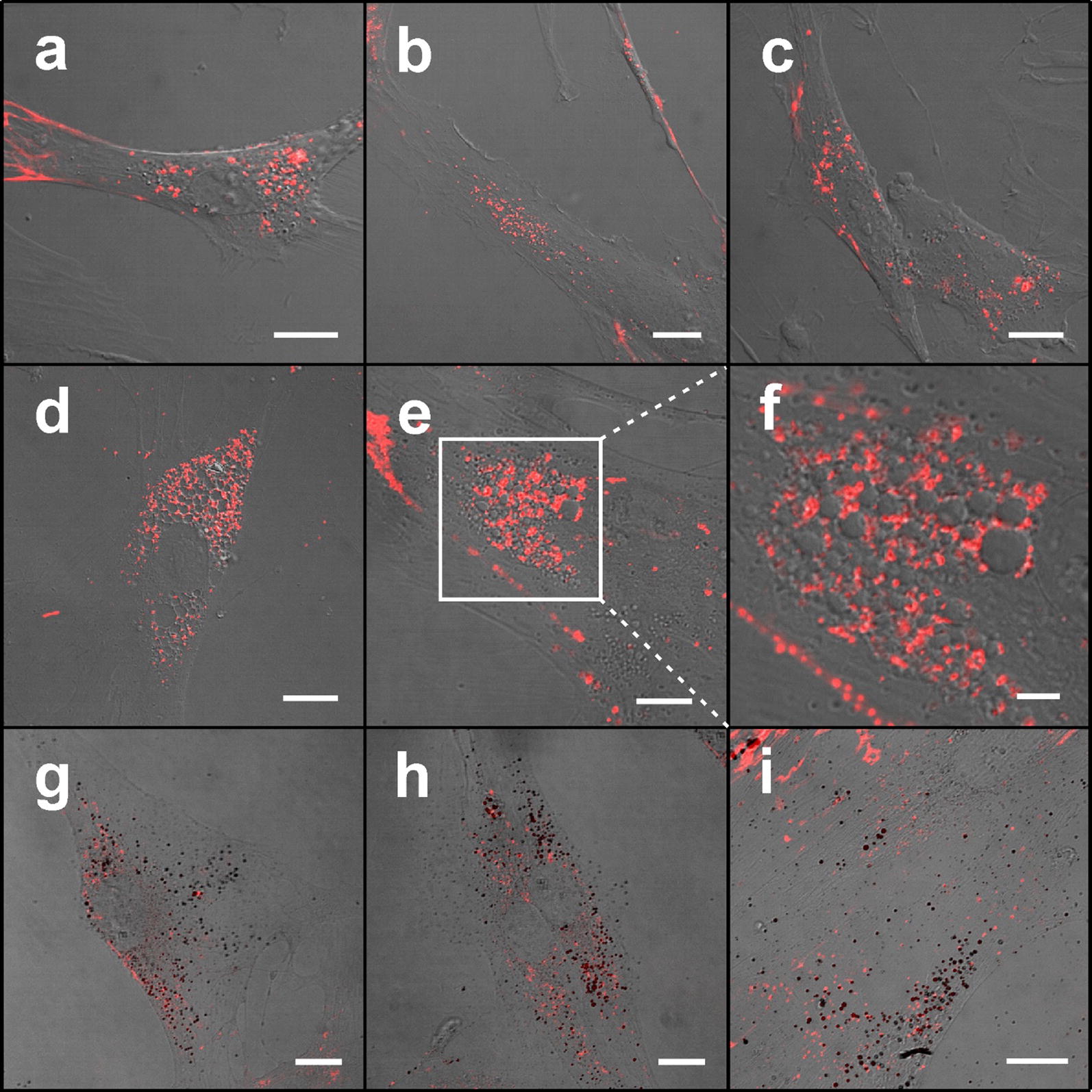
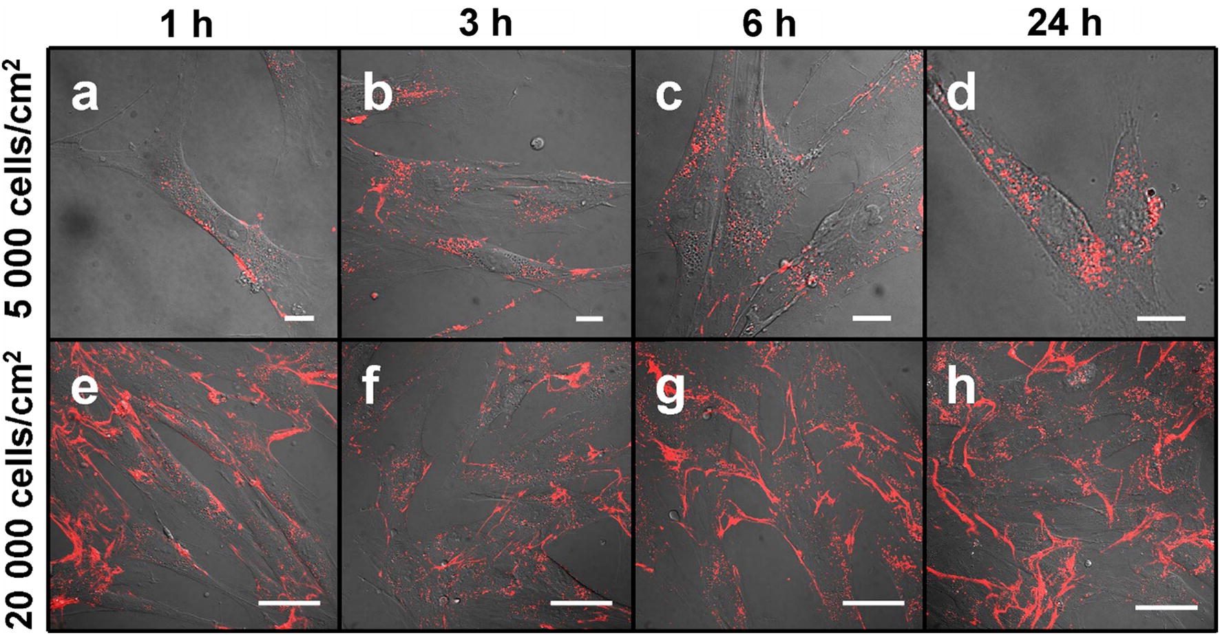
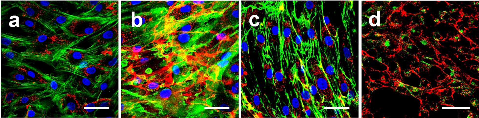
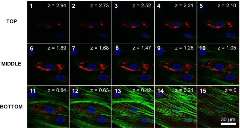
QDs had no negative impact on MSC viability throughout the experiment and accumulated in all observed cells efficiently; however, in some MSCs QDs induced formation of lipid droplets. At low cell growing densities QDs distribute within MSCs cytoplasm already after 1 h of incubation reaching saturation after 6 h. After 24 h QDs localize mainly in the perinuclear region of the cells in endosomes. Interestingly, in more confluent culture QDs localize mostly outside MSCs. QDs abundantly mark MSC long filopodia-like structures attaching neighboring cells. At high cell density cultivation, we for the first time demonstrated that carboxylated QDs localize in human bone marrow MSC extracellular matrix. Moreover, we observed that average photoluminescence lifetime of QDs distributed in extracellular matrix are longer than lifetimes of QDs entrapped in endocytic vesicles; thus, for the first time showing the possibility to identify and distinguish localization of QDs in various extracellular and intracellular structures using fluorescence-lifetime imaging microscopy without additional staining assays.

CONCLUSION

Carboxylated QDs can be used as nonspecific and effective dye for staining of human bone marrow MSCs and their specific extracellular structures. These results are promising in fundamental stem cell biology as well as in cellular therapy, anticancer drug delivery and tissue engineering.

摘要

背景

人类间充质干细胞(MSCs)因其免疫调节和抗炎作用而在再生医学领域引起了广泛关注。MSCs 具有特定的肿瘤靶向迁移和整合特性,这突出了 MSCs 作为抗癌药物理想载体的潜力。骨髓是临床应用中 MSCs 的主要来源。MSCs 的体内示踪是治疗性细胞产品安全性和疗效评估的关键组成部分;因此,细胞必须用对比剂进行标记,以实现 MSCs 体内迁移的可视化。由于其独特的性质,量子点(QDs)在长期 MSC 光学成像应用中成为最佳工具。本研究旨在研究非靶向羧基化量子点在不同细胞生长密度下在人骨髓间充质干细胞中的摄取动力学、细胞毒性、亚细胞和细胞外分布。

结果

在整个实验过程中,QDs 对 MSC 活力没有负面影响,并能有效地在所有观察到的细胞中积累;然而,在一些 MSC 中,QDs 诱导了脂滴的形成。在低细胞生长密度下,QDs 在孵育 1 小时后即可分布在 MSC 细胞质中,并在 6 小时后达到饱和。孵育 24 小时后,QDs 主要定位于细胞的核周区,位于内体中。有趣的是,在更致密的培养物中,QDs 主要位于 MSC 之外。QDs 大量标记 MSC 长丝状伪足状结构,附着相邻细胞。在高细胞密度培养时,我们首次证明了羧基化 QDs 定位于人骨髓 MSC 细胞外基质中。此外,我们观察到分布在细胞外基质中的 QDs 的平均荧光寿命长于内体中 QDs 的荧光寿命;因此,首次表明可以使用荧光寿命成像显微镜来识别和区分 QDs 在各种细胞外和细胞内结构中的定位,而无需额外的染色测定。

结论

羧基化 QDs 可用作非特异性和有效染料,用于染色人骨髓间充质干细胞及其特定的细胞外结构。这些结果在基础干细胞生物学以及细胞治疗、抗癌药物输送和组织工程中具有广阔的应用前景。

https://cdn.ncbi.nlm.nih.gov/pmc/blobs/7e85/6417192/1a73aa3d009b/12951_2019_470_Fig6_HTML.jpg
https://cdn.ncbi.nlm.nih.gov/pmc/blobs/7e85/6417192/9cd727f3438e/12951_2019_470_Fig1_HTML.jpg
https://cdn.ncbi.nlm.nih.gov/pmc/blobs/7e85/6417192/93dd597d9290/12951_2019_470_Fig2_HTML.jpg
https://cdn.ncbi.nlm.nih.gov/pmc/blobs/7e85/6417192/89a71be7cb89/12951_2019_470_Fig3_HTML.jpg
https://cdn.ncbi.nlm.nih.gov/pmc/blobs/7e85/6417192/bd7c01c1d9ae/12951_2019_470_Fig4_HTML.jpg
https://cdn.ncbi.nlm.nih.gov/pmc/blobs/7e85/6417192/6bac97338d09/12951_2019_470_Fig5_HTML.jpg
https://cdn.ncbi.nlm.nih.gov/pmc/blobs/7e85/6417192/1a73aa3d009b/12951_2019_470_Fig6_HTML.jpg

相似文献

1
Uptake and distribution of carboxylated quantum dots in human mesenchymal stem cells: cell growing density matters.羧基化量子点在人骨髓间充质干细胞中的摄取和分布:细胞生长密度很重要。
J Nanobiotechnology. 2019 Mar 13;17(1):39. doi: 10.1186/s12951-019-0470-6.
2
Polyamidoamine dendrimer-conjugated quantum dots for efficient labeling of primary cultured mesenchymal stem cells.聚酰胺-胺树枝状聚合物偶联量子点用于原代培养间充质干细胞的高效标记。
Biomaterials. 2011 Oct;32(28):6676-82. doi: 10.1016/j.biomaterials.2011.05.076. Epub 2011 Jun 22.
3
Skin-derived mesenchymal stem cells as quantum dot vehicles to tumors.皮肤来源的间充质干细胞作为肿瘤的量子点载体。
Int J Nanomedicine. 2017 Nov 6;12:8129-8142. doi: 10.2147/IJN.S143367. eCollection 2017.
4
Persistence of fluorescent nanoparticle-labelled bone marrow mesenchymal stem cells in vitro and after intra-articular injection.荧光纳米颗粒标记骨髓间充质干细胞在体外和关节内注射后的持久性。
J Tissue Eng Regen Med. 2019 Feb;13(2):191-202. doi: 10.1002/term.2781. Epub 2019 Jan 23.
5
Labeling and imaging mesenchymal stem cells with quantum dots.用量子点标记和成像间充质干细胞。
Methods Mol Biol. 2012;906:199-210. doi: 10.1007/978-1-61779-953-2_15.
6
The internalized CdSe/ZnS quantum dots impair the chondrogenesis of bone marrow mesenchymal stem cells.内化的CdSe/ZnS量子点损害骨髓间充质干细胞的软骨形成。
J Biomed Mater Res B Appl Biomater. 2006 Oct;79(1):95-101. doi: 10.1002/jbm.b.30517.
7
Functional quantum dot-siRNA nanoplexes to regulate chondrogenic differentiation of mesenchymal stem cells.用于调节间充质干细胞软骨分化的功能性量子点-siRNA纳米复合物
Acta Biomater. 2016 Dec;46:165-176. doi: 10.1016/j.actbio.2016.09.008. Epub 2016 Sep 9.
8
Fate of bone marrow mesenchymal stromal cells following autologous transplantation in a rabbit model of osteonecrosis.自体移植后骨髓间充质基质细胞在兔骨坏死模型中的命运
Cytotherapy. 2016 Feb;18(2):198-204. doi: 10.1016/j.jcyt.2015.10.016.
9
Nano-engineered skin mesenchymal stem cells: potential vehicles for tumour-targeted quantum-dot delivery.纳米工程化皮肤间充质干细胞:肿瘤靶向量子点递送的潜在载体
Beilstein J Nanotechnol. 2017 Jun 7;8:1218-1230. doi: 10.3762/bjnano.8.123. eCollection 2017.
10
Labeling of mesenchymal stem cells with bioconjugated quantum dots.用生物共轭量子点标记间充质干细胞。
Methods Mol Biol. 2011;680:61-75. doi: 10.1007/978-1-60761-901-7_4.

引用本文的文献

1
Simple and Cost-Effective Generation of 3D Cell Sheets and Spheroids Using Curvature-Controlled Paraffin Wax Substrates.使用曲率控制的石蜡基质简单且经济高效地生成3D细胞片和球体。
Nano Converg. 2024 Oct 31;11(1):44. doi: 10.1186/s40580-024-00451-4.
2
Pharmacokinetic characteristics of mesenchymal stem cells in translational challenges.间充质干细胞在转化挑战中的药代动力学特征。
Signal Transduct Target Ther. 2024 Sep 13;9(1):242. doi: 10.1038/s41392-024-01936-8.
3
Quantum Dots as a Potential Multifunctional Material for the Enhancement of Clinical Diagnosis Strategies and Cancer Treatments.

本文引用的文献

1
Silver nanoparticle based coatings enhance adipogenesis compared to osteogenesis in human mesenchymal stem cells through oxidative stress.基于银纳米颗粒的涂层通过氧化应激,与人骨髓间充质干细胞中的成骨作用相比,增强了脂肪生成。
J Mater Chem B. 2016 Feb 28;4(8):1466-1479. doi: 10.1039/c5tb02482j. Epub 2016 Feb 3.
2
Interaction of carboxylated CdSe/ZnS quantum dots with fish embryos: Towards understanding of nanoparticles toxicity.羧基化 CdSe/ZnS 量子点与鱼类胚胎的相互作用:探究纳米颗粒毒性。
Sci Total Environ. 2018 Sep 1;635:1280-1291. doi: 10.1016/j.scitotenv.2018.04.206. Epub 2018 Apr 24.
3
What exactly is 'N' in cell culture and animal experiments?
量子点作为一种潜在的多功能材料用于增强临床诊断策略和癌症治疗。
Nanomaterials (Basel). 2024 Jun 25;14(13):1088. doi: 10.3390/nano14131088.
4
Synthesis of metformin-derived fluorescent quantum dots: uptake, cytotoxicity, and inhibition in human breast cancer cells through autophagy pathway.二甲双胍衍生荧光量子点的合成:通过自噬途径在人乳腺癌细胞中的摄取、细胞毒性及抑制作用
J Biol Eng. 2024 Jun 25;18(1):38. doi: 10.1186/s13036-024-00433-4.
5
Recent advances of nanoparticles on bone tissue engineering and bone cells.纳米颗粒在骨组织工程和骨细胞方面的最新进展。
Nanoscale Adv. 2024 Feb 12;6(8):1957-1973. doi: 10.1039/d3na00851g. eCollection 2024 Apr 16.
6
In Vitro Tracking of Human Umbilical Vein Endothelial Cells Using Ultra-Sensitive Quantum Dot-Embedded Silica Nanoparticles.利用超灵敏量子点嵌入硅纳米粒子对人脐静脉内皮细胞进行体外跟踪。
Int J Mol Sci. 2023 Mar 17;24(6):5794. doi: 10.3390/ijms24065794.
7
Enhancing Stem Cell-Based Therapeutic Potential by Combining Various Bioengineering Technologies.通过结合多种生物工程技术提高基于干细胞的治疗潜力。
Front Cell Dev Biol. 2022 Jul 5;10:901661. doi: 10.3389/fcell.2022.901661. eCollection 2022.
8
Broad-Spectrum Theranostics and Biomedical Application of Functionalized Nanomaterials.功能化纳米材料的广谱诊疗学及生物医学应用
Polymers (Basel). 2022 Mar 17;14(6):1221. doi: 10.3390/polym14061221.
9
Virus-Mimicking Polymer Nanoparticles Targeting CD169 Macrophages as Long-Acting Nanocarriers for Combination Antiretrovirals.病毒模拟聚合物纳米颗粒靶向 CD169 巨噬细胞作为长效纳米载体用于联合抗逆转录病毒治疗。
ACS Appl Mater Interfaces. 2022 Jan 19;14(2):2488-2500. doi: 10.1021/acsami.1c17415. Epub 2022 Jan 7.
10
Bone Morphogenetic Protein-2 Conjugated to Quantum Dots is Biologically Functional.与量子点结合的骨形态发生蛋白-2具有生物学功能。
Nanomaterials (Basel). 2020 Jun 20;10(6):1208. doi: 10.3390/nano10061208.
细胞培养和动物实验中的“N”到底是什么?
PLoS Biol. 2018 Apr 4;16(4):e2005282. doi: 10.1371/journal.pbio.2005282. eCollection 2018 Apr.
4
Nanoparticle delivery to metastatic breast cancer cells by nanoengineered mesenchymal stem cells.纳米工程化间充质干细胞向转移性乳腺癌细胞递送纳米颗粒
Beilstein J Nanotechnol. 2018 Jan 29;9:321-332. doi: 10.3762/bjnano.9.32. eCollection 2018.
5
Superparamagnetic Iron Oxide-Loaded Cationic Polymersomes for Cellular MR Imaging of Therapeutic Stem Cells in Stroke.用于中风治疗性干细胞细胞磁共振成像的超顺磁性氧化铁负载阳离子聚合物囊泡
J Biomed Nanotechnol. 2016 Dec;12(12):2112-24. doi: 10.1166/jbn.2016.2321.
6
Skin-derived mesenchymal stem cells as quantum dot vehicles to tumors.皮肤来源的间充质干细胞作为肿瘤的量子点载体。
Int J Nanomedicine. 2017 Nov 6;12:8129-8142. doi: 10.2147/IJN.S143367. eCollection 2017.
7
Nano-engineered skin mesenchymal stem cells: potential vehicles for tumour-targeted quantum-dot delivery.纳米工程化皮肤间充质干细胞:肿瘤靶向量子点递送的潜在载体
Beilstein J Nanotechnol. 2017 Jun 7;8:1218-1230. doi: 10.3762/bjnano.8.123. eCollection 2017.
8
Use of Nanoparticle Contrast Agents for Cell Tracking with Computed Tomography.纳米颗粒造影剂在计算机断层扫描细胞追踪中的应用。
Bioconjug Chem. 2017 Jun 21;28(6):1581-1597. doi: 10.1021/acs.bioconjchem.7b00194. Epub 2017 May 18.
9
Usage of Human Mesenchymal Stem Cells in Cell-based Therapy: Advantages and Disadvantages.人间充质干细胞在细胞治疗中的应用:优点与缺点。
Dev Reprod. 2017 Mar;21(1):1-10. doi: 10.12717/DR.2017.21.1.001. Epub 2017 Mar 31.
10
Quick and effective method of bone marrow mesenchymal stem cell extraction.快速有效的骨髓间充质干细胞提取方法。
Open Med (Wars). 2014 Oct 8;10(1):44-49. doi: 10.1515/med-2015-0008. eCollection 2015.