Venkatesh Ramesh, Sinha Shivani, Gangadharaiah Deepika, Gadde Santosh G K, Mohan Ashwin, Shetty Rohit, Yadav Naresh Kumar
1Department of Retina and Vitreous, Narayana Nethralaya, #121/C, 1st R Block, Chord RoadRajaji Nagar, Bengaluru, 560010 India.
2Department of Cornea and Refractive surgery, Narayana Nethralaya, #121/C, 1st R Block, Chord Road, Rajaji Nagar, Bengaluru, 560010 India.
Eye Vis (Lond). 2019 Mar 7;6:8. doi: 10.1186/s40662-019-0133-6. eCollection 2019.
To examine the retinal structure-vascular-function relationship using optical coherence tomography (OCT) and optical coherence tomography angiography (OCTA) in myopia.
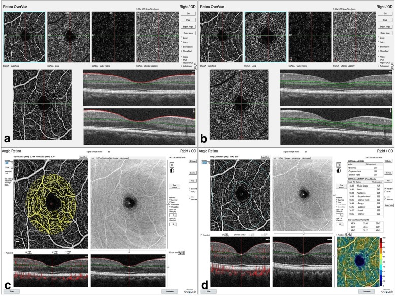
This was a prospective cross-sectional study comprising 86 eyes of 45 individuals with varying axial lengths and spherical equivalents and no posterior segment abnormalities. All eyes underwent optical coherence tomography with the Spectralis SD-OCT and OCTA with RTVue-XR Avanti; Optovue. Individual macular retinal layer thicknesses and flow areas and vessel densities were measured on OCT and OCTA, respectively. Linear correlations were made between the macular layer thicknesses, flow areas and vessel densities with axial length, spherical equivalent and visual acuity.
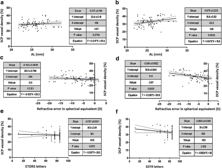
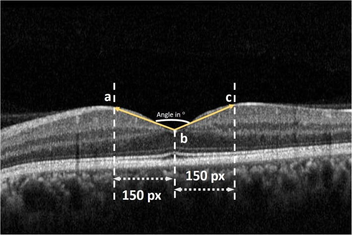
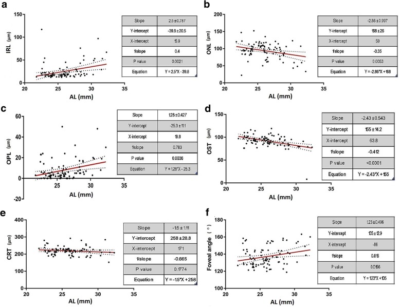
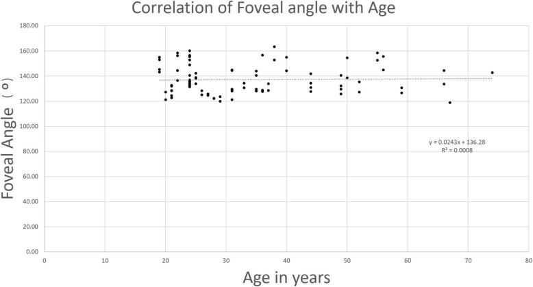
The participants' mean ages were 33.34 ± 14.45 years, mean spherical equivalent refractions were - 7.17 ± 5.71 D and axial lengths were 25.95 ± 2.41 mm. There were significant positive correlations of foveal angle (r = 0.757, = 0.001), inner retinal (r = 0.764, p = 0.001) and outer plexiform layer (r = 0.771, p = 0.001) thickness on OCT and vessel densities in deep capillary plexus (r = 0.313, = 0.003) on OCTA with axial length and negative correlations with spherical equivalents and visual acuity. Significant negative correlations of outer nuclear layer (r = - 0.560, = 0.03) and photoreceptor outer segment layer thickness (r = - 0.856, < 0.001) were noted on OCT with axial length and positive correlations with spherical equivalents and visual acuity.
The lateral retinal stretching in myopia could possibly explain the correlation between retinal layer thickness, vascular density and visual acuity in these eyes. Further research is required to investigate this.
使用光学相干断层扫描(OCT)和光学相干断层扫描血管造影(OCTA)研究近视眼中视网膜结构-血管-功能的关系。
这是一项前瞻性横断面研究,纳入了45名个体的86只眼,这些个体具有不同的眼轴长度和球镜等效度数,且无眼后段异常。所有眼睛均使用Spectralis SD-OCT进行光学相干断层扫描,并使用RTVue-XR Avanti;Optovue进行OCTA检查。分别在OCT和OCTA上测量个体黄斑视网膜各层厚度、血流面积和血管密度。对黄斑层厚度、血流面积和血管密度与眼轴长度、球镜等效度数和视力进行线性相关分析。
参与者的平均年龄为33.34±14.45岁,平均球镜等效屈光度为-7.17±5.71D,眼轴长度为25.95±2.41mm。OCT上的黄斑中心凹角度(r = 0.757,p = 0.001)、视网膜内层(r = 0.764,p = 0.001)和外丛状层(r = 0.771,p = 0.001)厚度与OCTA上深层毛细血管丛的血管密度(r = 0.313,p = 0.003)与眼轴长度呈显著正相关,与球镜等效度数和视力呈负相关。OCT上外核层(r = -0.560,p = 0.03)和光感受器外段层厚度(r = -0.856,p < 0.001)与眼轴长度呈显著负相关,与球镜等效度数和视力呈正相关。
近视眼中视网膜的横向拉伸可能解释了这些眼中视网膜层厚度、血管密度和视力之间的相关性。需要进一步研究对此进行调查。