• 文献检索
  • 文档翻译
  • 深度研究
  • 学术资讯
  • Suppr Zotero 插件Zotero 插件
  • 邀请有礼
  • 套餐&价格
  • 历史记录
应用&插件
Suppr Zotero 插件Zotero 插件浏览器插件Mac 客户端Windows 客户端微信小程序
定价
高级版会员购买积分包购买API积分包
服务
文献检索文档翻译深度研究API 文档MCP 服务
关于我们
关于 Suppr公司介绍联系我们用户协议隐私条款
关注我们

Suppr 超能文献

核心技术专利:CN118964589B侵权必究
粤ICP备2023148730 号-1Suppr @ 2026

文献检索

告别复杂PubMed语法,用中文像聊天一样搜索,搜遍4000万医学文献。AI智能推荐,让科研检索更轻松。

立即免费搜索

文件翻译

保留排版,准确专业,支持PDF/Word/PPT等文件格式,支持 12+语言互译。

免费翻译文档

深度研究

AI帮你快速写综述,25分钟生成高质量综述,智能提取关键信息,辅助科研写作。

立即免费体验

近视眼中黄斑区血管密度降低。

Reduced Macular Vascular Density in Myopic Eyes.

作者信息

Fan Hua, Chen Hao-Yu, Ma Hong-Jie, Chang Zheng, Yin Hai-Quan, Ng Danny Siu-Chun, Cheung Carol Y, Hu Shan, Xiang Xiang, Tang Shi-Bo, Li Shuang-Nong

机构信息

Aier School of Ophthalmology, Central South University, Changsha, Hunan 410015, China.

Joint Shantou International Eye Center, Shantou, Guangdong 515000, China.

出版信息

Chin Med J (Engl). 2017 Feb 20;130(4):445-451. doi: 10.4103/0366-6999.199844.

DOI:10.4103/0366-6999.199844
PMID:28218219
原文链接:https://pmc.ncbi.nlm.nih.gov/articles/PMC5324382/
Abstract

BACKGROUND

Morphological changes of the vasculature system in patients with myopia have been observed by Doppler ultrasound and fundus fluorescein angiography (FFA); however, these studies have limitations. Doppler ultrasound provides low-resolution images which are mainly obtained from visualized large vessels, and FFA is an invasive examination. Optic coherence tomography (OCT) angiography is a noninvasive, high-resolution measurement for vascular density. The purpose of this study was to investigate the change of vascular density in myopic eyes using OCT angiography.

METHODS

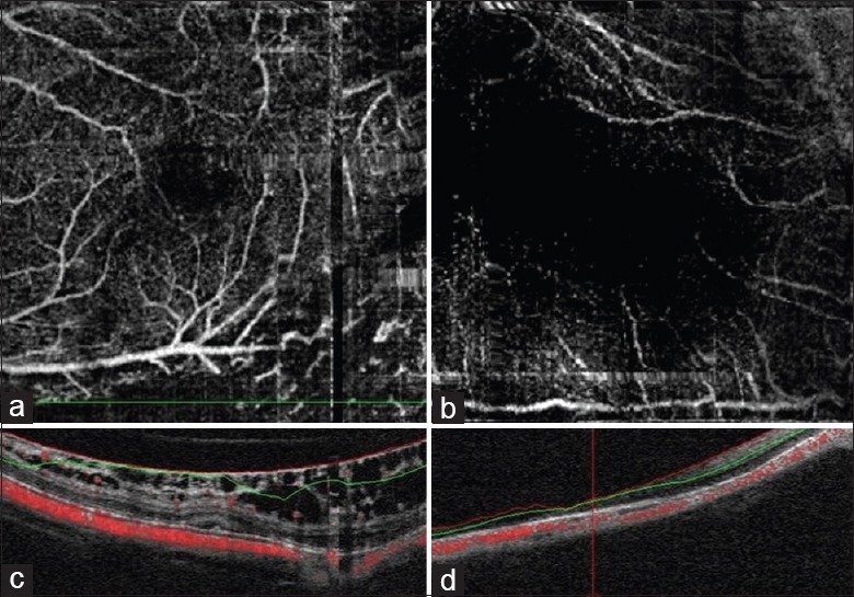
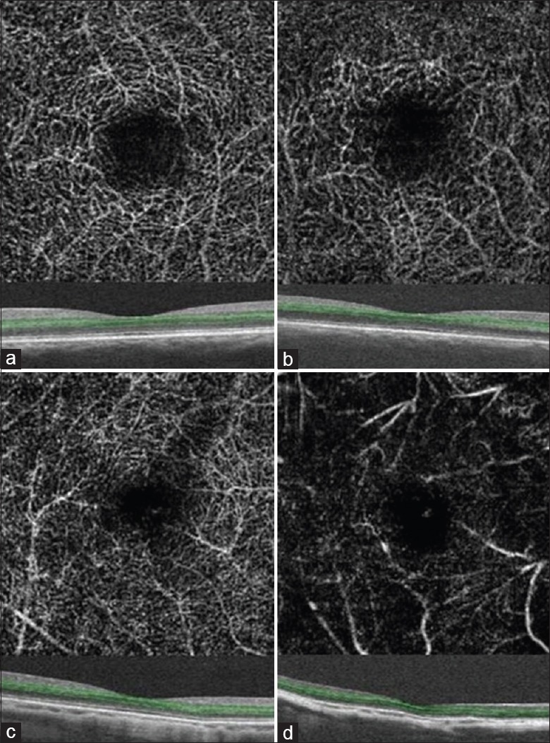
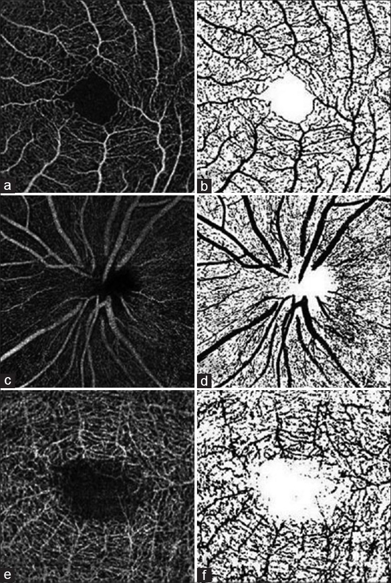
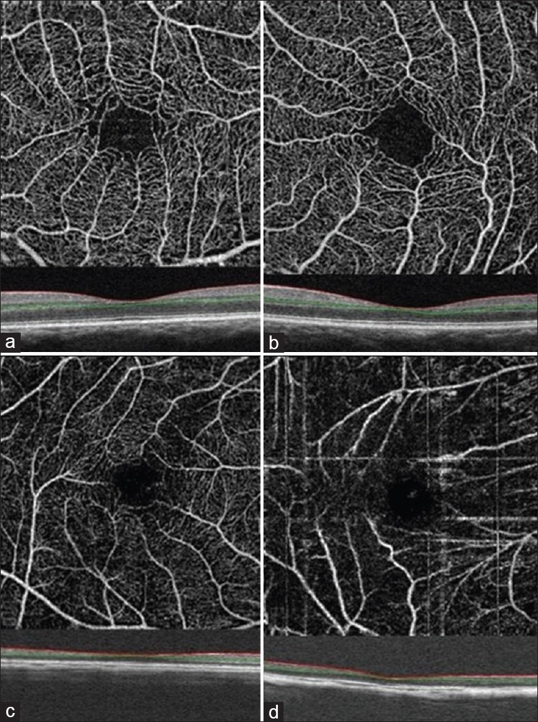
This cross-sectional study includes a total of 91 eyes from 47 participants including control, moderate, and high myopia that were evaluated by OCT angiography. Patients with myopia were recruited from the Refractive Department, Shenzhen Aier Eye Hospital, from August 5, 2015 to April 1, 2016. Emmetropic eyes were from healthy volunteers. The vascular density at macula and optic disc regions, ganglion cell complex (GCC) thickness, and retinal nerve fiber layer (RNFL) thickness were measured. Their relationships with axial length (AL) and refractive error were analyzed. One-way analysis of variance (ANOVA), Pearson's correlation, and generalized estimating equation were used for statistical analysis.

RESULTS

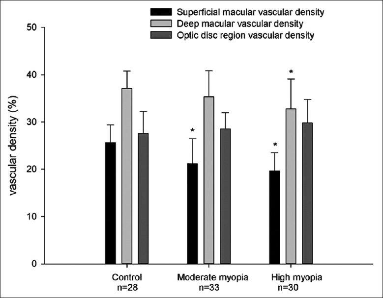
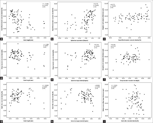
Both superficial and deep macular vascular density were highest in control (25.64% ± 3.76% and 37.12% ± 3.66%, respectively), then in moderate myopia (21.15% ± 5.33% and 35.35% ± 5.50%, respectively), and lowest in high myopia group (19.64% ± 3.87% and 32.81% ± 6.29%, respectively) (F = 13.74 and 4.57, respectively; both P < 0.001). Both superficial (β = -0.850 and 0.460, respectively) and deep (β = -0.766 and 0.396, respectively) macular vascular density were associated with AL and spherical equivalent (all P < 0.001). Superficial macular vascular density was associated with GCC thickness (β = 0.244, P = 0.040), independent of spherical equivalent. The vascular density in optic disc region had no difference among the three groups, and it was not associated with AL, spherical equivalent, or RNFL thickness.

CONCLUSION

Our results suggested that with the increase of myopia, the vascular density decreased in macular region, but not in optic disc region.

摘要

背景

通过多普勒超声和眼底荧光血管造影(FFA)已观察到近视患者血管系统的形态学变化;然而,这些研究存在局限性。多普勒超声提供的是低分辨率图像,主要从可视化的大血管获取,而FFA是一种侵入性检查。光学相干断层扫描(OCT)血管造影是一种用于血管密度的非侵入性、高分辨率测量方法。本研究的目的是使用OCT血管造影研究近视眼血管密度的变化。

方法

这项横断面研究共纳入了47名参与者的91只眼睛,包括对照组、中度近视组和高度近视组,均通过OCT血管造影进行评估。2015年8月5日至2016年4月1日期间,从深圳爱尔眼科医院屈光科招募近视患者。正视眼来自健康志愿者。测量黄斑和视盘区域的血管密度、神经节细胞复合体(GCC)厚度以及视网膜神经纤维层(RNFL)厚度。分析它们与眼轴长度(AL)和屈光不正的关系。采用单因素方差分析(ANOVA)、Pearson相关性分析和广义估计方程进行统计分析。

结果

对照组黄斑浅层和深层血管密度最高(分别为25.64%±3.76%和37.12%±3.66%),其次是中度近视组(分别为21.15%±5.33%和35.35%±5.50%),高度近视组最低(分别为19.64%±3.87%和32.81%±6.29%)(F分别为13.74和4.57;P均<0.001)。黄斑浅层(β分别为-0.850和0.460)和深层(β分别为-0.766和0.396)血管密度均与眼轴长度和等效球镜相关(P均<0.001)。黄斑浅层血管密度与GCC厚度相关(β = 0.244;P = 0.040),独立于等效球镜。视盘区域血管密度在三组之间无差异,且与眼轴长度、等效球镜或RNFL厚度均无关。

结论

我们的结果表明,随着近视程度增加,黄斑区域血管密度降低,但视盘区域未降低。

https://cdn.ncbi.nlm.nih.gov/pmc/blobs/cb29/5324382/033d06e6e1a1/CMJ-130-445-g006.jpg
https://cdn.ncbi.nlm.nih.gov/pmc/blobs/cb29/5324382/6c648c2021a3/CMJ-130-445-g001.jpg
https://cdn.ncbi.nlm.nih.gov/pmc/blobs/cb29/5324382/c7a1b6acef14/CMJ-130-445-g002.jpg
https://cdn.ncbi.nlm.nih.gov/pmc/blobs/cb29/5324382/8432590806f4/CMJ-130-445-g003.jpg
https://cdn.ncbi.nlm.nih.gov/pmc/blobs/cb29/5324382/0d4c2e5a2cb5/CMJ-130-445-g004.jpg
https://cdn.ncbi.nlm.nih.gov/pmc/blobs/cb29/5324382/1d5eab6ca09b/CMJ-130-445-g005.jpg
https://cdn.ncbi.nlm.nih.gov/pmc/blobs/cb29/5324382/033d06e6e1a1/CMJ-130-445-g006.jpg

相似文献

1
Reduced Macular Vascular Density in Myopic Eyes.近视眼中黄斑区血管密度降低。
Chin Med J (Engl). 2017 Feb 20;130(4):445-451. doi: 10.4103/0366-6999.199844.
2
Ganglion cell-inner plexiform layer and retinal nerve fiber layer thickness according to myopia and optic disc area: a quantitative and three-dimensional analysis.根据近视和视盘面积分析神经节细胞-内网状层及视网膜神经纤维层厚度:定量与三维分析
BMC Ophthalmol. 2017 Mar 11;17(1):22. doi: 10.1186/s12886-017-0419-1.
3
Vascular flow density in pathological myopia: an optical coherence tomography angiography study.病理性近视中的血管血流密度:一项光学相干断层扫描血管造影研究。
BMJ Open. 2017 Feb 3;7(2):e013571. doi: 10.1136/bmjopen-2016-013571.
4
Myopic optic disc tilt and the characteristics of peripapillary retinal nerve fiber layer thickness measured by spectral-domain optical coherence tomography.高度近视视盘倾斜与频域光学相干断层扫描测量的视盘周围视网膜神经纤维层厚度的特征。
J Glaucoma. 2012 Apr-May;21(4):260-5. doi: 10.1097/IJG.0b013e31820719e1.
5
Influence of Myopia on Size of Optic Nerve Head and Retinal Nerve Fiber Layer Thickness Measured by Spectral Domain Optical Coherence Tomography.近视对通过频域光学相干断层扫描测量的视神经乳头大小和视网膜神经纤维层厚度的影响。
Korean J Ophthalmol. 2016 Oct;30(5):335-343. doi: 10.3341/kjo.2016.30.5.335. Epub 2016 Sep 29.
6
Evaluation of retinal vascular density and related factors in youth myopia without maculopathy using OCTA.使用光学相干断层扫描血管造影(OCTA)评估无黄斑病变的青少年近视患者的视网膜血管密度及相关因素。
Sci Rep. 2021 Jul 28;11(1):15361. doi: 10.1038/s41598-021-94909-8.
7
[Peripapillary and macular vessel density in eyes with different phases of thyroid-associated ophthalmopathy].[甲状腺相关眼病不同阶段眼睛的视乳头周围和黄斑区血管密度]
Zhonghua Yan Ke Za Zhi. 2020 Nov 11;56(11):824-831. doi: 10.3760/cma.j.cn112142-20191115-00574.
8
Assessment of superficial retinal microvascular density in healthy myopia.健康近视患者视网膜浅层微血管密度的评估
Int Ophthalmol. 2019 Aug;39(8):1861-1870. doi: 10.1007/s10792-018-1014-z. Epub 2018 Sep 3.
9
[Macular and retinal nerve fiber layer thickness in myopic children by three-dimensional OCT].[三维光学相干断层扫描测量近视儿童黄斑和视网膜神经纤维层厚度]
Zhonghua Yi Xue Za Zhi. 2013 Dec 3;93(45):3573-6.
10
Impact of Valsalva maneuver on central choroid, central macula, and disk fiber layer thickness among high myopic and hyperopic patients.瓦氏动作对高度近视和远视患者中央脉络膜、中央黄斑及视盘纤维层厚度的影响。
Eur J Ophthalmol. 2017 May 11;27(3):331-335. doi: 10.5301/ejo.5000834. Epub 2016 Jul 18.

引用本文的文献

1
Differences in macular structure and microvasculature between high myopia and non-high myopia.高度近视与非高度近视之间黄斑结构和微血管系统的差异。
Front Med (Lausanne). 2025 Aug 26;12:1645015. doi: 10.3389/fmed.2025.1645015. eCollection 2025.
2
Effect of Refractive Status on Peripapillary Retinal Nerve Fiber Layer Thickness and Vascular Density: A Prospective Optical Coherence Tomography Angiography-Based Study.屈光状态对视乳头周围视网膜神经纤维层厚度和血管密度的影响:一项基于光学相干断层扫描血管造影的前瞻性研究。
Med J Islam Repub Iran. 2025 Feb 5;39:21. doi: 10.47176/mjiri.39.21. eCollection 2025.
3
Interocular comparison of peripapillary retinal nerve fiber layer thickness and vasculature in non-pathological myopia with anisometropia.

本文引用的文献

1
A Novel Tectonic Keratoplasty with Femtosecond Laser Intrastromal Lenticule for Corneal Ulcer and Perforation.一种用于角膜溃疡和穿孔的新型飞秒激光基质内透镜植入式角膜移植术。
Chin Med J (Engl). 2016 Aug 5;129(15):1817-21. doi: 10.4103/0366-6999.186639.
2
Reproducibility of Perfusion Parameters of Optic Disc and Macula in Rhesus Monkeys by Optical Coherence Tomography Angiography.光学相干断层扫描血管造影术测量恒河猴视盘和黄斑灌注参数的可重复性
Chin Med J (Engl). 2016 May 5;129(9):1087-90. doi: 10.4103/0366-6999.180532.
3
Advances of optical coherence tomography in myopia and pathologic myopia.
非病理性屈光参差性近视患者视乳头周围视网膜神经纤维层厚度和血管系统的双眼比较
Graefes Arch Clin Exp Ophthalmol. 2025 Apr 8. doi: 10.1007/s00417-025-06826-5.
4
The Significance of OCTA in Studying Vessel Density and Retinal Thickness in Individuals with Myopia.光学相干断层扫描血管造影(OCTA)在研究近视患者血管密度和视网膜厚度中的意义
Medicina (Kaunas). 2025 Mar 18;61(3):532. doi: 10.3390/medicina61030532.
5
Sustained Experimental Myopia Exacerbates the Effect of Eye Growth on Retinal Ganglion Cell Density and Function.持续性实验性近视会加剧眼球生长对视网膜神经节细胞密度和功能的影响。
Int J Mol Sci. 2025 Mar 20;26(6):2824. doi: 10.3390/ijms26062824.
6
Relationship Between Axial Length and Retinal Oxygen Dynamics in Adults With Myopia.成人近视患者眼轴长度与视网膜氧动力学的关系
Transl Vis Sci Technol. 2025 Jan 2;14(1):18. doi: 10.1167/tvst.14.1.18.
7
Compartmental analysis of retinal vascular parameters and thickness in myopic eyes using SS-OCTA.使用扫频光学相干断层扫描血管造影(SS-OCTA)对近视眼视网膜血管参数和厚度进行分区分析。
Front Med (Lausanne). 2024 Dec 20;11:1521710. doi: 10.3389/fmed.2024.1521710. eCollection 2024.
8
Evaluation of retinal and choroidal microvascular alteration after ICL V4c implantation based on OCTA.基于 OCTA 评价 ICL V4c 植入术后视网膜及脉络膜微血管改变。
BMC Ophthalmol. 2024 Nov 6;24(1):487. doi: 10.1186/s12886-024-03752-8.
9
Structural, blood flow and functional changes in the macular area and the expression of aqueous humor factors in myopia.近视患者黄斑区的结构、血流及功能变化以及房水因子的表达
Front Med (Lausanne). 2024 Jul 17;11:1335084. doi: 10.3389/fmed.2024.1335084. eCollection 2024.
10
Correlation in retinal thickness and macular retina plus choroidal microcirculation in pediatric myopia.儿童近视的视网膜厚度与黄斑视网膜加脉络膜微循环的相关性。
Medicine (Baltimore). 2024 Jul 26;103(30):e38915. doi: 10.1097/MD.0000000000038915.
光学相干断层扫描在近视和病理性近视中的研究进展
Eye (Lond). 2016 Jul;30(7):901-16. doi: 10.1038/eye.2016.47. Epub 2016 Apr 8.
4
Is the peripapillary retinal perfusion related to myopia in healthy eyes? A prospective comparative study.健康眼中视乳头周围视网膜灌注与近视有关吗?一项前瞻性对照研究。
BMJ Open. 2016 Mar 11;6(3):e010791. doi: 10.1136/bmjopen-2015-010791.
5
Longitudinal and Cross-Sectional Analyses of Age Effects on Retinal Nerve Fiber Layer and Ganglion Cell Complex Thickness by Fourier-Domain OCT.傅里叶域光学相干断层扫描对年龄对视网膜神经纤维层和神经节细胞复合体厚度影响的纵向和横断面分析
Transl Vis Sci Technol. 2016 Mar 4;5(2):1. doi: 10.1167/tvst.5.2.1. eCollection 2016 Mar.
6
In Vivo Assessment of Macular Vascular Density in Healthy Human Eyes Using Optical Coherence Tomography Angiography.使用光学相干断层扫描血管造影术对健康人眼黄斑血管密度进行体内评估。
Am J Ophthalmol. 2016 May;165:39-46. doi: 10.1016/j.ajo.2016.02.018. Epub 2016 Feb 24.
7
IMAGE ARTIFACTS IN OPTICAL COHERENCE TOMOGRAPHY ANGIOGRAPHY.光学相干断层扫描血管造影中的图像伪影
Retina. 2015 Nov;35(11):2163-80. doi: 10.1097/IAE.0000000000000765.
8
Retinal vessel oxygen saturation and vessel diameter in high myopia.高度近视患者的视网膜血管血氧饱和度及血管直径
Ophthalmic Physiol Opt. 2015 Sep;35(5):562-9. doi: 10.1111/opo.12223.
9
Correlation between optic disc perfusion and glaucomatous severity in patients with open-angle glaucoma: an optical coherence tomography angiography study.开角型青光眼患者视盘灌注与青光眼严重程度的相关性:一项光学相干断层扫描血管造影研究。
Graefes Arch Clin Exp Ophthalmol. 2015 Sep;253(9):1557-64. doi: 10.1007/s00417-015-3095-y. Epub 2015 Aug 11.
10
Single-site Baseline and Short-term Outcomes of Clinical Characteristics and Life Quality Evaluation of Chinese Wet Age-related Macular Degeneration Patients in Routine Clinical Practice.中国湿性年龄相关性黄斑变性患者在常规临床实践中的单中心基线及短期临床特征和生活质量评估结果
Chin Med J (Engl). 2015 May 5;128(9):1154-9. doi: 10.4103/0366-6999.156083.