Department of Radiology, Mayo Clinic College of Medicine and Science, Mayo Clinic, Rochester, MN, USA.
Department of Urology, Mayo Clinic College of Medicine and Science, Mayo Clinic, Rochester, MN, USA.
Sci Rep. 2019 Mar 20;9(1):4907. doi: 10.1038/s41598-019-41373-0.
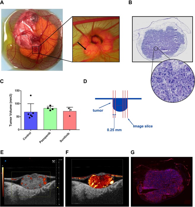
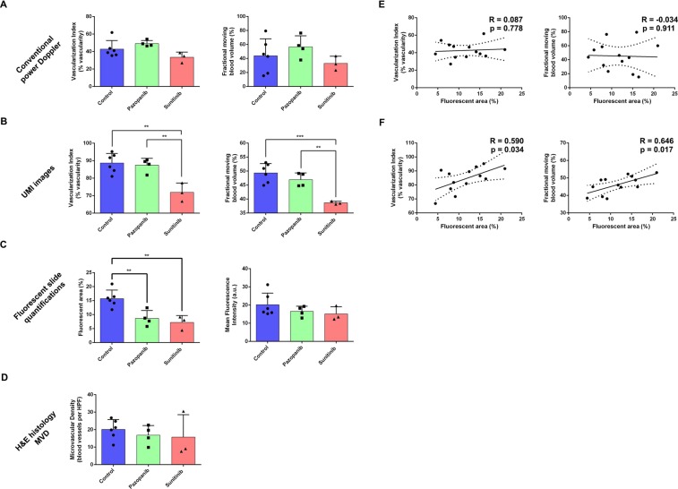
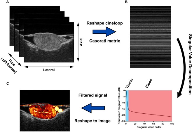
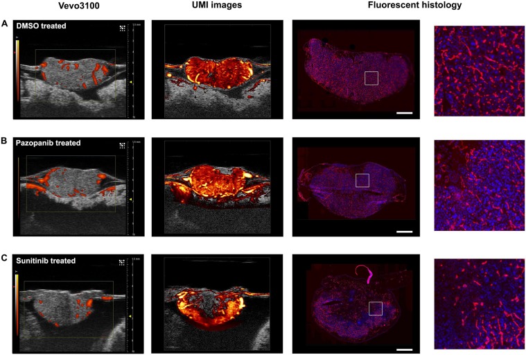
Ultrasound microvessel imaging (UMI), when applied with ultrafast planewave acquisitions, has demonstrated superior blood signal sensitivity in comparison to conventional Doppler imaging. Here we propose a high spatial resolution and ultra-sensitive UMI that is based on conventional line-by-line high-frequency ultrasound imagers and singular value decomposition (SVD) clutter filtering for the visualization and quantification of tumor microvasculature and perfusion. The technique was applied to a chicken embryo tumor model of renal cell carcinoma that was treated with two FDA-approved anti-angiogenic agents at clinically relevant dosages. We demonstrate the feasibility of 3D evaluation with UMI to achieve highly sensitive detection of microvasculature using conventional line-by-line ultrasound imaging on a preclinical and commercially available high-frequency ultrasound device without software or hardware modifications. Quantitative parameters (vascularization index and fractional moving blood volume) derived from UMI images provide significantly improved evaluation of anti-angiogenic therapy response as compared with conventional power Doppler imaging, using histological analysis and immunohistochemistry as the reference standard. This proof-of-concept study demonstrates that high-frequency UMI is a low-cost, contrast-agent-free, easily applicable, accessible, and quantitative imaging tool for tumor characterization, which may be very useful for preclinical evaluation and longitudinal monitoring of anti-cancer treatment.
超声微血管成像(UMI)与超快速平面波采集结合使用时,与传统的多普勒成像相比,具有更高的血液信号灵敏度。在这里,我们提出了一种基于传统逐线高频超声成像和奇异值分解(SVD)杂波滤波的高空间分辨率和超高灵敏度 UMI,用于可视化和量化肿瘤微血管和灌注。该技术应用于肾细胞癌鸡胚肿瘤模型,该模型用两种经 FDA 批准的抗血管生成剂以临床相关剂量进行治疗。我们证明了使用 UMI 进行 3D 评估的可行性,无需软件或硬件修改,即可使用常规逐线超声成像在临床前和商业上可获得的高频超声设备上实现对微血管的高灵敏度检测。与传统的功率多普勒成像相比,UMI 图像得出的定量参数(血管化指数和分数运动血容量)可显著改善抗血管生成治疗反应的评估,以组织学分析和免疫组织化学为参考标准。这项概念验证研究表明,高频 UMI 是一种低成本、无造影剂、易于应用、易于获得的定量成像工具,可用于肿瘤特征描述,对于癌症治疗的临床前评估和纵向监测可能非常有用。