Yukawa Yasutsugu, Matsumoto Taro, Kollor Heiko, Minamide Akihito, Hashizume Hiroshi, Yamada Hiroshi, Kato Fumihiko
Department of Orthopaedic Surgery, Wakayama Medical University, Wakayama, Japan.
Department of Orthopaedic Surgery, Chubu Rosai Hospital, Nagoya, Japan.
Asian Spine J. 2019 Mar 26;13(4):663-671. doi: 10.31616/asj.2018.0187. Print 2019 Aug.
Prospective cohort imaging study.
This study aimed to evaluate lumbar sagittal alignment and range of motion (ROM) using radiographs in a large asymptomatic cohort and identify sex-based differences and age-related changes in the subjects.
Several researchers have tried to establish normal alignment and kinematic behavior of the lumbar spine, using plain radiographs. Few studies have employed a large and sex-and age-balanced cohort.
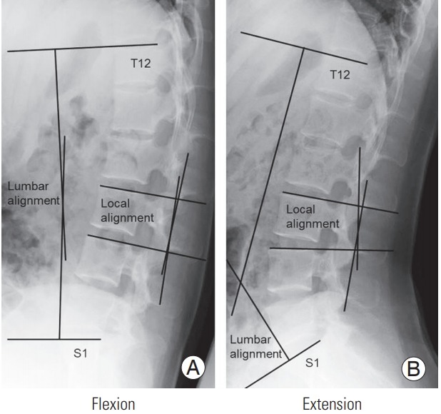
Total 627 healthy volunteers (at least 50 males and 50 females in each age decade, from the 3rd to the 8th decade) underwent whole spine radiography in the standing position; lumbar spine radiography was performed for all subjects in the recumbent position. Lumbar lordosis (LL, T12-S1) and ROM during flexion and extension were measured using a computer digitizer.
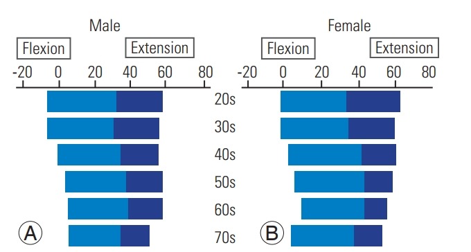
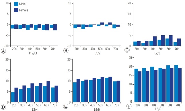
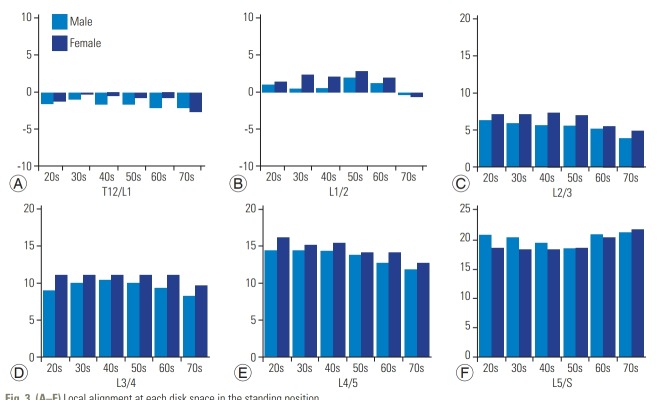
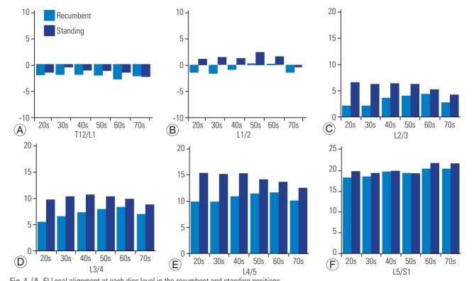
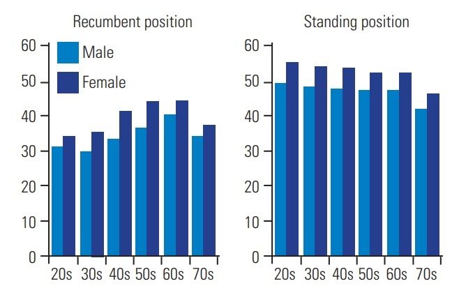
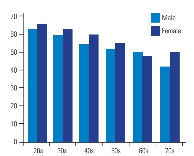
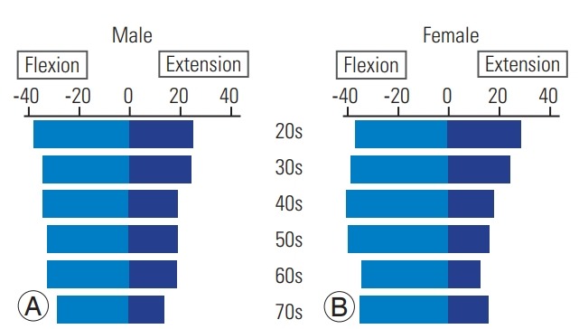
The mean LL was 36.8°±13.2° in the recumbent position and 49.8°±11.2° in the standing position. The LL was greater in the standing position than in the recumbent position; further, LL was higher in females as compared to that in males. Local lordosis at each disk level increased incrementally with distal progression through the lumbar spine in both the positions. Local lordosis at L4- S1 was 29.8°±8.0° in the recumbent position and 34.2°±8.3° in the standing position and occupied 85.1% and 70.8% of the total LL, respectively. However, local lordosis in the standing position decreased with age at L2-3, L3-4, and L4-5 levels. Total lumbar ROM (T12-S1) decreased with age. The ROM in females was higher than that in males.
We established the standard value and age-related changes in the lumbar alignment and ROM in each age decade in asymptomatic subjects. These data will be useful and provide the normal values for comparison in clinical practice to identify sexbased differences and age-related changes.
前瞻性队列影像学研究。
本研究旨在通过X线片评估一大群无症状人群的腰椎矢状面排列和活动范围(ROM),并确定受试者基于性别的差异和与年龄相关的变化。
几位研究人员试图利用普通X线片确定腰椎的正常排列和运动学行为。很少有研究采用大规模且性别和年龄均衡的队列。
总共627名健康志愿者(每个年龄十年至少50名男性和50名女性,年龄范围从30岁到80岁)站立位进行全脊柱X线摄影;所有受试者卧位进行腰椎X线摄影。使用计算机数字化仪测量腰椎前凸(LL,T12-S1)以及屈伸过程中的ROM。
卧位时平均LL为36.8°±13.2°,站立位时为49.8°±11.2°。站立位时的LL大于卧位时;此外,女性的LL高于男性。在两个体位中,每个椎间盘水平的局部前凸均随着腰椎向远端进展而逐渐增加。L4-S1节段卧位时局部前凸为29.8°±8.0°,站立位时为34.2°±8.3°,分别占总LL的85.1%和70.8%。然而,在L2-3、L3-4和L4-5节段,站立位时的局部前凸随年龄增长而降低。腰椎总ROM(T12-S1)随年龄增长而降低。女性的ROM高于男性。
我们确定了无症状受试者各年龄十年腰椎排列和ROM的标准值以及与年龄相关的变化。这些数据将有助于临床实践中进行比较,以识别基于性别的差异和与年龄相关的变化,并提供正常参考值。