• 文献检索
  • 文档翻译
  • 深度研究
  • 学术资讯
  • Suppr Zotero 插件Zotero 插件
  • 邀请有礼
  • 套餐&价格
  • 历史记录
应用&插件
Suppr Zotero 插件Zotero 插件浏览器插件Mac 客户端Windows 客户端微信小程序
定价
高级版会员购买积分包购买API积分包
服务
文献检索文档翻译深度研究API 文档MCP 服务
关于我们
关于 Suppr公司介绍联系我们用户协议隐私条款
关注我们

Suppr 超能文献

核心技术专利:CN118964589B侵权必究
粤ICP备2023148730 号-1Suppr @ 2026

文献检索

告别复杂PubMed语法,用中文像聊天一样搜索,搜遍4000万医学文献。AI智能推荐,让科研检索更轻松。

立即免费搜索

文件翻译

保留排版,准确专业,支持PDF/Word/PPT等文件格式,支持 12+语言互译。

免费翻译文档

深度研究

AI帮你快速写综述,25分钟生成高质量综述,智能提取关键信息,辅助科研写作。

立即免费体验

影像组学特征和多层感知机网络分类器:用于鉴别胶质母细胞瘤和原发性中枢神经系统淋巴瘤的稳健 MRI 分类策略。

Radiomic features and multilayer perceptron network classifier: a robust MRI classification strategy for distinguishing glioblastoma from primary central nervous system lymphoma.

机构信息

Department of Convergence Medicine, University of Ulsan College of Medicine, Asan Medical Center, 88 Olympic-Ro 43-Gil Songpa-Gu, Seoul, 05505, Korea.

Department of Radiology and Research Institute of Radiology, University of Ulsan College of Medicine, 88 Olympic-Ro 43-Gil Songpa-Gu, Seoul, 05505, Korea.

出版信息

Sci Rep. 2019 Apr 5;9(1):5746. doi: 10.1038/s41598-019-42276-w.

DOI:10.1038/s41598-019-42276-w
PMID:30952930
原文链接:https://pmc.ncbi.nlm.nih.gov/articles/PMC6451024/
Abstract

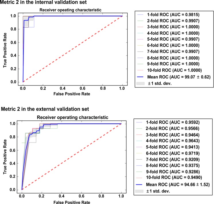
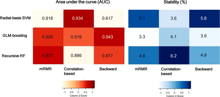
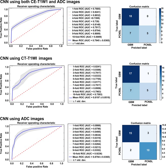
We aimed to establish a high-performing and robust classification strategy, using magnetic resonance imaging (MRI), along with combinations of feature extraction and selection in human and machine learning using radiomics or deep features by employing a small dataset. Using diffusion and contrast-enhanced T1-weighted MR images obtained from patients with glioblastomas and primary central nervous system lymphomas, classification task was assigned to a combination of radiomic features and (1) supervised machine learning after feature selection or (2) multilayer perceptron (MLP) network; or MR image input without radiomic feature extraction to (3) two neuro-radiologists or (4) an end-to-end convolutional neural network (CNN). The results showed similar high performance in generalized linear model (GLM) classifier and MLP using radiomics features in the internal validation set, but MLP network remained robust in the external validation set obtained using different MRI protocols. CNN showed the lowest performance in both validation sets. Our results reveal that a combination of radiomic features and MLP network classifier serves a high-performing and generalizable model for classification task for a small dataset with heterogeneous MRI protocols.

摘要

我们旨在建立一种高性能且稳健的分类策略,使用磁共振成像(MRI),结合放射组学或深度学习特征的特征提取和选择,在人类和机器学习中使用小数据集。使用从患有胶质母细胞瘤和原发性中枢神经系统淋巴瘤的患者获得的弥散和对比增强 T1 加权 MR 图像,分类任务被分配给放射组学特征的组合和(1)经过特征选择的监督机器学习,或(2)多层感知机(MLP)网络;或不进行放射组学特征提取的 MR 图像输入到(3)两位神经放射科医生或(4)端到端卷积神经网络(CNN)。结果表明,在内部验证集中,使用放射组学特征的广义线性模型(GLM)分类器和 MLP 具有相似的高性能,但 MLP 网络在使用不同 MRI 协议获得的外部验证集中仍然稳健。CNN 在两个验证集中的表现均最低。我们的结果表明,放射组学特征和 MLP 网络分类器的组合为具有异构 MRI 协议的小数据集的分类任务提供了一种高性能且可推广的模型。

https://cdn.ncbi.nlm.nih.gov/pmc/blobs/252b/6451024/1b717fea9299/41598_2019_42276_Fig4_HTML.jpg
https://cdn.ncbi.nlm.nih.gov/pmc/blobs/252b/6451024/29c95fe56ba9/41598_2019_42276_Fig1_HTML.jpg
https://cdn.ncbi.nlm.nih.gov/pmc/blobs/252b/6451024/57c0a0acd7b6/41598_2019_42276_Fig2_HTML.jpg
https://cdn.ncbi.nlm.nih.gov/pmc/blobs/252b/6451024/0c0513001712/41598_2019_42276_Fig3_HTML.jpg
https://cdn.ncbi.nlm.nih.gov/pmc/blobs/252b/6451024/1b717fea9299/41598_2019_42276_Fig4_HTML.jpg

相似文献

1
Radiomic features and multilayer perceptron network classifier: a robust MRI classification strategy for distinguishing glioblastoma from primary central nervous system lymphoma.影像组学特征和多层感知机网络分类器:用于鉴别胶质母细胞瘤和原发性中枢神经系统淋巴瘤的稳健 MRI 分类策略。
Sci Rep. 2019 Apr 5;9(1):5746. doi: 10.1038/s41598-019-42276-w.
2
Radiomics features to distinguish glioblastoma from primary central nervous system lymphoma on multi-parametric MRI.基于多参数MRI的区分胶质母细胞瘤与原发性中枢神经系统淋巴瘤的影像组学特征
Neuroradiology. 2018 Dec;60(12):1297-1305. doi: 10.1007/s00234-018-2091-4. Epub 2018 Sep 19.
3
Machine Learning-based Texture Analysis of Contrast-enhanced MR Imaging to Differentiate between Glioblastoma and Primary Central Nervous System Lymphoma.基于机器学习的对比增强磁共振成像纹理分析鉴别胶质母细胞瘤和原发性中枢神经系统淋巴瘤。
Magn Reson Med Sci. 2019 Jan 10;18(1):44-52. doi: 10.2463/mrms.mp.2017-0178. Epub 2018 May 16.
4
Glioblastoma and primary central nervous system lymphoma: differentiation using MRI derived first-order texture analysis - a machine learning study.胶质母细胞瘤和原发性中枢神经系统淋巴瘤:基于 MRI 一阶纹理分析的鉴别——一项机器学习研究。
Neuroradiol J. 2021 Aug;34(4):320-328. doi: 10.1177/1971400921998979. Epub 2021 Mar 3.
5
Diffusion radiomics as a diagnostic model for atypical manifestation of primary central nervous system lymphoma: development and multicenter external validation.扩散放射组学作为原发性中枢神经系统淋巴瘤不典型表现的诊断模型:开发和多中心外部验证。
Neuro Oncol. 2018 Aug 2;20(9):1251-1261. doi: 10.1093/neuonc/noy021.
6
Primary central nervous system lymphoma and atypical glioblastoma: Differentiation using radiomics approach.原发性中枢神经系统淋巴瘤和非典型性脑胶质瘤:基于放射组学方法的鉴别诊断。
Eur Radiol. 2018 Sep;28(9):3832-3839. doi: 10.1007/s00330-018-5368-4. Epub 2018 Apr 6.
7
Application of MR morphologic, diffusion tensor, and perfusion imaging in the classification of brain tumors using machine learning scheme.基于机器学习方案的磁共振形态学、弥散张量和灌注成像在脑肿瘤分类中的应用。
Neuroradiology. 2019 Jul;61(7):757-765. doi: 10.1007/s00234-019-02195-z. Epub 2019 Apr 4.
8
Robust performance of deep learning for distinguishing glioblastoma from single brain metastasis using radiomic features: model development and validation.深度学习利用放射组学特征区分胶质母细胞瘤与单发脑转移瘤的稳健性能:模型的建立与验证。
Sci Rep. 2020 Jul 21;10(1):12110. doi: 10.1038/s41598-020-68980-6.
9
Classification of glioblastoma versus primary central nervous system lymphoma using convolutional neural networks.基于卷积神经网络的胶质母细胞瘤与原发性中枢神经系统淋巴瘤的分类。
Sci Rep. 2021 Jul 26;11(1):15219. doi: 10.1038/s41598-021-94733-0.
10
Radiomics-based differentiation between glioblastoma and primary central nervous system lymphoma: a comparison of diagnostic performance across different MRI sequences and machine learning techniques.基于放射组学的脑胶质母细胞瘤与原发性中枢神经系统淋巴瘤鉴别:不同 MRI 序列和机器学习技术的诊断效能比较。
Eur Radiol. 2021 Nov;31(11):8703-8713. doi: 10.1007/s00330-021-07845-6. Epub 2021 Apr 23.

引用本文的文献

1
AI-Driven Innovations in Neuroradiology and Neurosurgery: Scoping Review of Current Evidence and Future Directions.人工智能驱动的神经放射学与神经外科创新:当前证据与未来方向的范围综述
Cancers (Basel). 2025 Aug 11;17(16):2625. doi: 10.3390/cancers17162625.
2
Radiomics and Clinical Features for Distinguishing Kidney Stone-Associated Urinary Tract Infection: A Comprehensive Analysis of Machine Learning Classification.用于区分肾结石相关性尿路感染的影像组学和临床特征:机器学习分类的综合分析
Open Forum Infect Dis. 2024 Oct 5;11(10):ofae581. doi: 10.1093/ofid/ofae581. eCollection 2024 Oct.
3
PET radiomics in lung cancer: advances and translational challenges.

本文引用的文献

1
The Relative Performance of Ensemble Methods with Deep Convolutional Neural Networks for Image Classification.深度卷积神经网络集成方法在图像分类中的相对性能
J Appl Stat. 2018;45(15):2800-2818. doi: 10.1080/02664763.2018.1441383. Epub 2018 Feb 26.
2
Diffusion radiomics as a diagnostic model for atypical manifestation of primary central nervous system lymphoma: development and multicenter external validation.扩散放射组学作为原发性中枢神经系统淋巴瘤不典型表现的诊断模型:开发和多中心外部验证。
Neuro Oncol. 2018 Aug 2;20(9):1251-1261. doi: 10.1093/neuonc/noy021.
3
Radiomics: the bridge between medical imaging and personalized medicine.
肺癌中的PET放射组学:进展与转化挑战
EJNMMI Phys. 2024 Oct 3;11(1):81. doi: 10.1186/s40658-024-00685-5.
4
Beyond hand-crafted features for pretherapeutic molecular status identification of pediatric low-grade gliomas.超越手工特征,用于小儿低级别胶质瘤治疗前分子状态的识别。
Sci Rep. 2024 Aug 17;14(1):19102. doi: 10.1038/s41598-024-69870-x.
5
Exploring the Potential of Machine Learning Algorithms to Improve Diffusion Nuclear Magnetic Resonance Imaging Models Analysis.探索机器学习算法在改善扩散核磁共振成像模型分析方面的潜力。
J Med Phys. 2024 Apr-Jun;49(2):189-202. doi: 10.4103/jmp.jmp_10_24. Epub 2024 Jun 25.
6
Improving the Generalizability and Performance of an Ultrasound Deep Learning Model Using Limited Multicenter Data for Lung Sliding Artifact Identification.利用有限的多中心数据提高用于肺滑动伪像识别的超声深度学习模型的通用性和性能
Diagnostics (Basel). 2024 May 22;14(11):1081. doi: 10.3390/diagnostics14111081.
7
Distinguishing diffuse large B-cell lymphoma from Hodgkin's lymphoma in children using an enhanced computed tomography radiomics approach.使用增强计算机断层扫描影像组学方法区分儿童弥漫性大B细胞淋巴瘤与霍奇金淋巴瘤。
Transl Pediatr. 2024 May 31;13(5):716-726. doi: 10.21037/tp-23-586. Epub 2024 May 28.
8
Fully automated MR-based virtual biopsy of primary CNS lymphomas.基于磁共振成像的原发性中枢神经系统淋巴瘤全自动虚拟活检
Neurooncol Adv. 2024 Mar 14;6(1):vdae022. doi: 10.1093/noajnl/vdae022. eCollection 2024 Jan-Dec.
9
Radiomics for differentiation of gliomas from primary central nervous system lymphomas: a systematic review and meta-analysis.基于影像组学鉴别胶质瘤与原发性中枢神经系统淋巴瘤的系统评价与Meta分析
Front Oncol. 2024 Feb 14;14:1291861. doi: 10.3389/fonc.2024.1291861. eCollection 2024.
10
Biomaterial-based 3D modeling of glioblastoma multiforme.基于生物材料的多形性胶质母细胞瘤3D建模。
Cancer Pathog Ther. 2023 Jan 9;1(3):177-194. doi: 10.1016/j.cpt.2023.01.002. eCollection 2023 Jul.
放射组学:医学影像与个性化医疗之间的桥梁。
Nat Rev Clin Oncol. 2017 Dec;14(12):749-762. doi: 10.1038/nrclinonc.2017.141. Epub 2017 Oct 4.
4
Lost in Thought - The Limits of the Human Mind and the Future of Medicine.陷入沉思——人类思维的局限与医学的未来
N Engl J Med. 2017 Sep 28;377(13):1209-1211. doi: 10.1056/NEJMp1705348.
5
A Deep Learning-Based Radiomics Model for Prediction of Survival in Glioblastoma Multiforme.基于深度学习的胶质母细胞瘤生存预测放射组学模型。
Sci Rep. 2017 Sep 4;7(1):10353. doi: 10.1038/s41598-017-10649-8.
6
Deep Learning based Radiomics (DLR) and its usage in noninvasive IDH1 prediction for low grade glioma.基于深度学习的放射组学(DLR)及其在低级别胶质瘤无创 IDH1 预测中的应用。
Sci Rep. 2017 Jul 14;7(1):5467. doi: 10.1038/s41598-017-05848-2.
7
Deep Learning in Medical Imaging: General Overview.医学成像中的深度学习:概述
Korean J Radiol. 2017 Jul-Aug;18(4):570-584. doi: 10.3348/kjr.2017.18.4.570. Epub 2017 May 19.
8
Deep Learning at Chest Radiography: Automated Classification of Pulmonary Tuberculosis by Using Convolutional Neural Networks.胸部放射摄影中的深度学习:使用卷积神经网络自动分类肺结核。
Radiology. 2017 Aug;284(2):574-582. doi: 10.1148/radiol.2017162326. Epub 2017 Apr 24.
9
Radiomics-based Prognosis Analysis for Non-Small Cell Lung Cancer.基于放射组学的非小细胞肺癌预后分析。
Sci Rep. 2017 Apr 18;7:46349. doi: 10.1038/srep46349.
10
MRI features predict survival and molecular markers in diffuse lower-grade gliomas.磁共振成像特征可预测弥漫性低级别胶质瘤的生存率及分子标志物。
Neuro Oncol. 2017 Jun 1;19(6):862-870. doi: 10.1093/neuonc/now256.