Department of Surgery, University of California, Los Angeles, 10833 Le Conte Ave, Los Angeles, CA, 90095, USA.
Department of Molecular and Medical Pharmacology, Crump Institute for Molecular Imaging, University of California, Los Angeles, CA, 90095, USA.
J Transl Med. 2019 Apr 5;17(1):113. doi: 10.1186/s12967-019-1862-y.
Various proinflammatory cytokines can be detected within the melanoma tumor microenvironment. Interleukin 32 (IL32) is produced by T cells, NK cells and monocytes/macrophages, but also by a subset of melanoma cells. We sought to better understand the biology of IL32 in human melanoma.
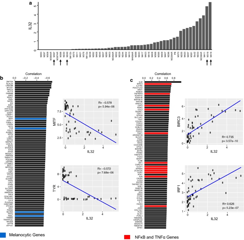
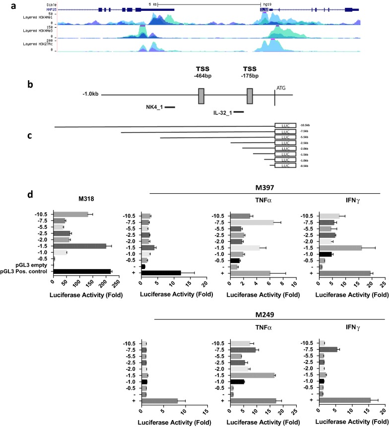
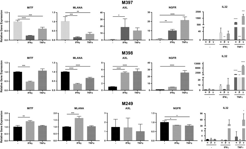
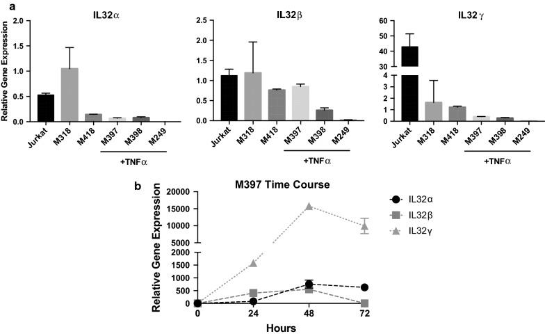
We analyzed RNA sequencing data from 53 in-house established human melanoma cell lines and 479 melanoma tumors from The Cancer Genome Atlas dataset. We evaluated global gene expression patterns associated with IL32 expression. We also evaluated the impact of proinflammatory molecules TNFα and IFNγ on IL32 expression and dedifferentiation in melanoma cell lines in vitro. In order to study the transcriptional regulation of IL32 in these cell lines, we cloned up to 10.5 kb of the 5' upstream region of the human IL32 gene into a luciferase reporter vector.
A significant proportion of established human melanoma cell lines express IL32, with its expression being highly correlated with a dedifferentiation genetic signature (high AXL/low MITF). Non IL32-expressing differentiated melanoma cell lines exposed to TNFα or IFNγ can be induced to express the three predominant isoforms (α, β, γ) of IL32. Cis-acting elements within this 5' upstream region of the human IL32 gene appear to govern both induced and constitutive gene expression. In the tumor microenvironment, IL32 expression is highly correlated with genes related to T cell infiltration, and also positively correlates with high AXL/low MITF dedifferentiated gene signature.
Expression of IL32 in human melanoma can be induced by TNFα or IFNγ and correlates with a treatment-resistant dedifferentiated genetic signature. Constitutive and induced expression are regulated, in part, by cis-acting sequences within the 5' upstream region.
多种促炎细胞因子可在黑色素瘤肿瘤微环境中检测到。白细胞介素 32(IL32)由 T 细胞、NK 细胞和单核细胞/巨噬细胞产生,但也由一部分黑色素瘤细胞产生。我们试图更好地了解 IL32 在人类黑色素瘤中的生物学特性。
我们分析了 53 个内部建立的人类黑色素瘤细胞系和来自癌症基因组图谱数据集的 479 个黑色素瘤肿瘤的 RNA 测序数据。我们评估了与 IL32 表达相关的全局基因表达模式。我们还评估了促炎分子 TNFα 和 IFNγ 对黑色素瘤细胞系中 IL32 表达和去分化的影响。为了研究这些细胞系中 IL32 的转录调控,我们将人类 IL32 基因的 5'上游区域克隆到荧光素酶报告载体中,长度可达 10.5 kb。
相当一部分已建立的人类黑色素瘤细胞系表达 IL32,其表达与去分化遗传特征(高 AXL/低 MITF)高度相关。非表达 IL32 的分化黑色素瘤细胞系暴露于 TNFα 或 IFNγ 可诱导其表达三种主要的同工型(α、β、γ)。人类 IL32 基因的这个 5'上游区域内的顺式作用元件似乎控制着诱导和组成型基因表达。在肿瘤微环境中,IL32 的表达与与 T 细胞浸润相关的基因高度相关,并且与高 AXL/低 MITF 去分化基因特征呈正相关。
人类黑色素瘤中 IL32 的表达可被 TNFα 或 IFNγ 诱导,并与治疗抵抗的去分化遗传特征相关。组成型和诱导型表达部分受 5'上游区域内的顺式作用序列调控。