Fukuda Yasunari, Asaoka Tadafumi, Eguchi Hidetoshi, Honma Keiichiro, Morii Eiichi, Iwagami Yoshifumi, Akita Hirofumi, Noda Takehiro, Gotoh Kunihito, Kobayashi Shogo, Mori Masaki, Doki Yuichiro
Department of Gastroenterological Surgery, Graduate School of Medicine, Osaka University, 2-2 Yamadaoka E-2, Suita, Osaka, 565-0871, Japan.
Department of Pathology, Graduate School of Medicine, Osaka University, Suita, Osaka, Japan.
Surg Case Rep. 2019 Apr 11;5(1):58. doi: 10.1186/s40792-019-0621-x.
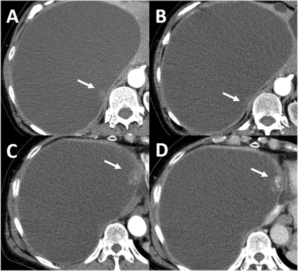
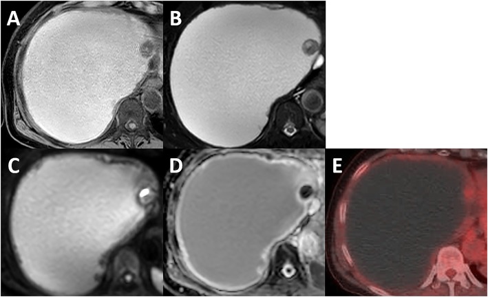
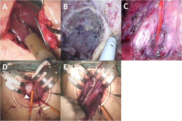
Hemorrhagic hepatic cysts infrequently involve several iconographic changes requiring a differential diagnosis, primarily with a cystic malignancy. We herein report a case of laparoscopy-assisted extended right hepatectomy for a giant hemorrhagic hepatic cyst with an enhancing mural nodule that was clinically suspected of being biliary cystadenocarcinoma.
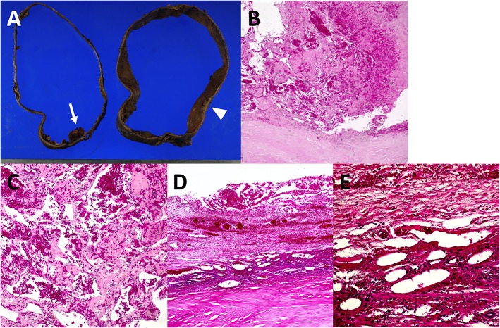
A 73-year-old woman was followed up for giant hepatic cyst occupying the right lobe of the liver. During the follow-up, an enhancing mural nodule was newly noted on computed tomography in 2016. Based on additional clinical examinations, biliary cystadenocarcinoma was undeniable, and laparoscopy-assisted extended right hepatectomy was performed for diagnostic and therapeutic purposes. She had no perioperative complications and was discharged on postoperative day 13. A histological examination of the mural nodule showed neovascularization within an organized hematoma.
We herein report a rare case of giant hemorrhagic hepatic cyst mimicking biliary cystadenocarcinoma that was successfully treated with laparoscopy-assisted extended right hepatectomy. Laparoscopic surgery in our case was an effective procedure performed with the utmost care.
出血性肝囊肿很少出现需要鉴别诊断的多种影像学改变,主要是与囊性恶性肿瘤鉴别。我们在此报告一例腹腔镜辅助扩大右肝切除术治疗巨大出血性肝囊肿的病例,该囊肿有一个强化的壁结节,临床上怀疑为胆管囊腺癌。
一名73岁女性因巨大肝囊肿占据肝脏右叶而接受随访。随访期间,2016年计算机断层扫描新发现一个强化的壁结节。基于进一步的临床检查,胆管囊腺癌难以排除,遂行腹腔镜辅助扩大右肝切除术以明确诊断并进行治疗。她没有围手术期并发症,术后第13天出院。壁结节的组织学检查显示在机化血肿内有新生血管形成。
我们在此报告一例罕见的巨大出血性肝囊肿酷似胆管囊腺癌的病例,该病例通过腹腔镜辅助扩大右肝切除术成功治疗。我们病例中的腹腔镜手术是一项极其谨慎进行的有效手术。