Ryu Sang Baek, Werginz Paul, Fried Shelley I
Boston VA Healthcare System, Boston, MA, United States.
Department of Neurosurgery, Massachusetts General Hospital, Harvard Medical School, Boston, MA, United States.
Front Neurosci. 2019 Apr 4;13:324. doi: 10.3389/fnins.2019.00324. eCollection 2019.
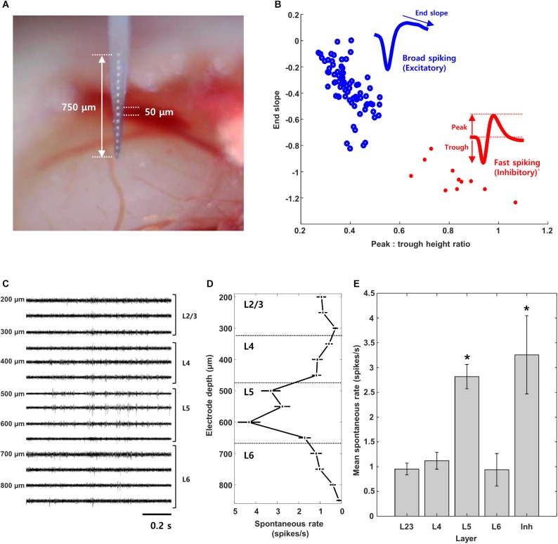
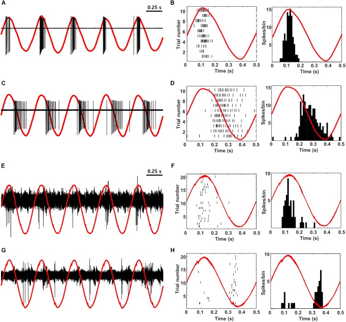
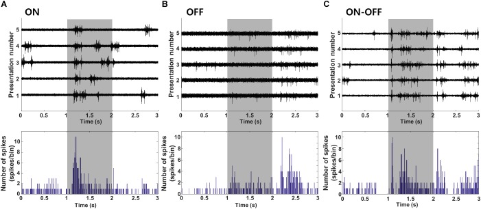
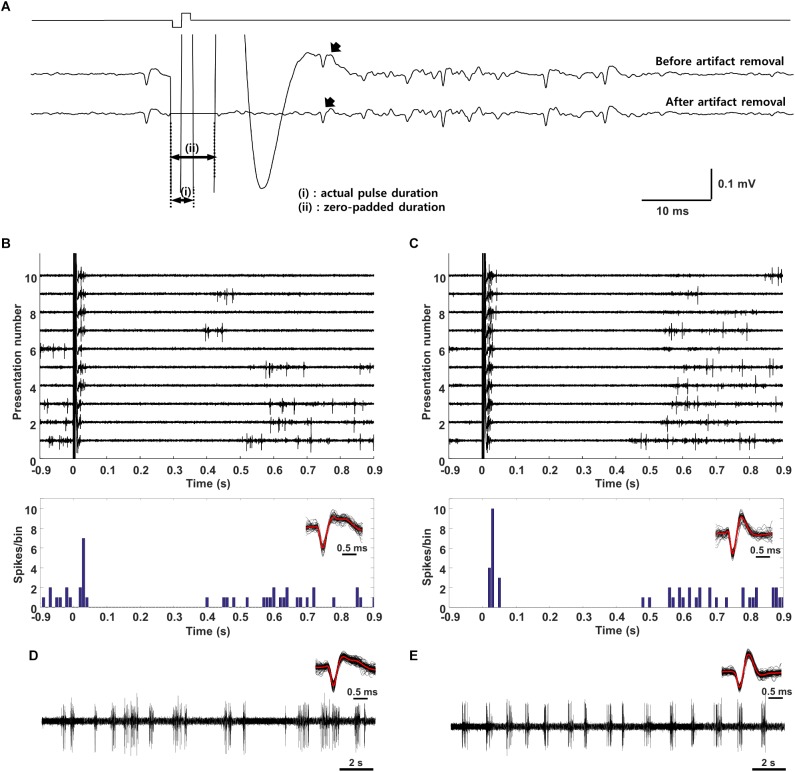
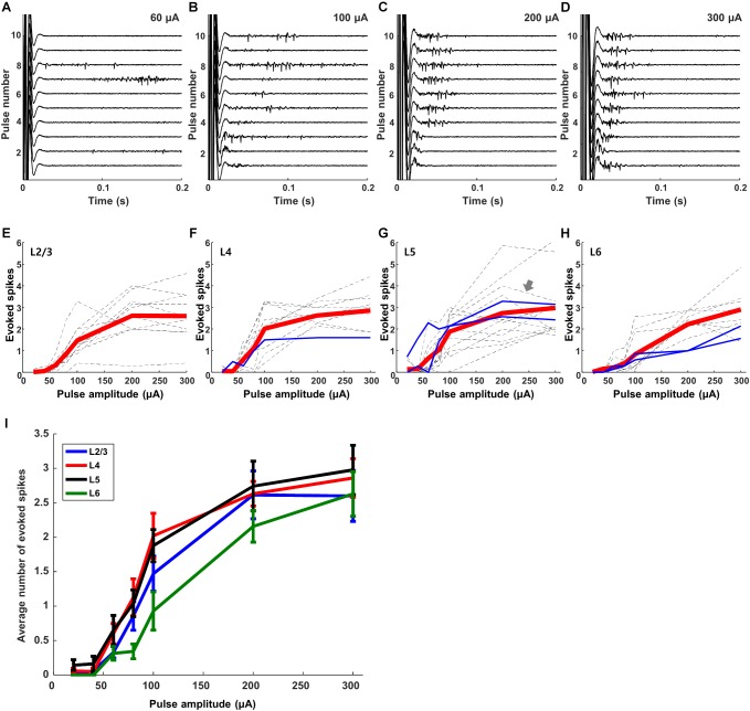
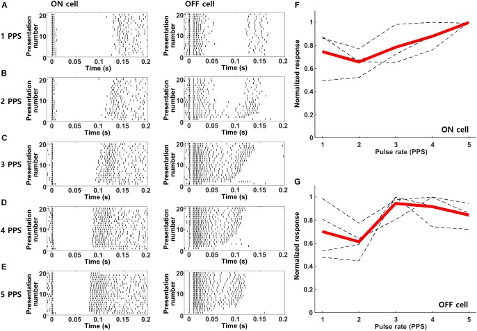
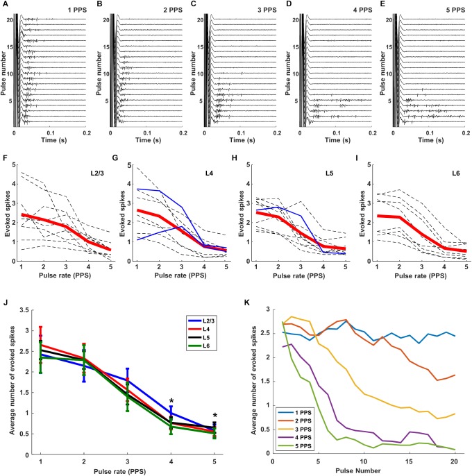
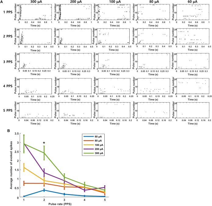
Retinal prostheses strive to restore vision to the blind by electrically stimulating the neurons that survive the disease process. Clinical effectiveness has been limited however, and much ongoing effort is devoted toward the development of improved stimulation strategies, especially ones that better replicate physiological patterns of neural signaling. Here, to better understand the potential effectiveness of different stimulation strategies, we explore the responses of neurons in the primary visual cortex to electric stimulation of the retina. A 16-channel implantable microprobe was used to record single unit activities from each layer of the mouse visual cortex. Layers were identified by electrode depth as well as spontaneous rate. Cell types were classified as excitatory or inhibitory based on their spike waveform and as ON, OFF, or ON-OFF based on the polarity of their light response. After classification, electric stimulation was delivered via a wire electrode placed on the surface of cornea (extraocularly) and responses were recorded from the cortex contralateral to the stimulated eye. Responses to electric stimulation were highly similar across cell types and layers. Responses (spike counts) increased as a function of the amplitude of stimulation, and although there was some variance across cells, the sensitivity to amplitude was largely similar across all cell types. Suppression of responses was observed for pulse rates ≥3 pulses per second (PPS) but did not originate in the retina as RGC responses remained stable to rates up to 5 PPS. Low-frequency sinusoids delivered to the retina replicated the out-of-phase responses that occur naturally in ON vs. OFF RGCs. Intriguingly, out-of-phase signaling persisted in V1 neurons, suggesting key aspects of neural signaling are preserved during transmission along visual pathways. Our results describe an approach to evaluate responses of cortical neurons to electric stimulation of the retina. By examining the responses of single cells, we were able to show that some retinal stimulation strategies can indeed better match the neural signaling patterns used by the healthy visual system. Because cortical signaling is better correlated to psychophysical percepts, the ability to evaluate which strategies produce physiological-like cortical responses may help to facilitate better clinical outcomes.
视网膜假体致力于通过电刺激在疾病过程中存活下来的神经元来恢复盲人的视力。然而,临床效果有限,目前许多工作都致力于开发改进的刺激策略,尤其是那些能更好地复制神经信号生理模式的策略。在此,为了更好地理解不同刺激策略的潜在效果,我们探索了初级视觉皮层中的神经元对视网膜电刺激的反应。使用一个16通道的可植入微探针记录小鼠视觉皮层各层的单个单元活动。通过电极深度以及自发发放率来识别各层。根据细胞的动作电位波形将细胞类型分为兴奋性或抑制性,并根据其光反应的极性分为ON、OFF或ON - OFF型。分类后,通过放置在角膜表面(眼外)的线电极进行电刺激,并记录受刺激眼对侧皮层的反应。不同细胞类型和层对电刺激的反应高度相似。反应(动作电位计数)随刺激幅度的增加而增加,尽管不同细胞之间存在一些差异,但所有细胞类型对幅度的敏感性在很大程度上是相似的。当脉冲频率≥3脉冲每秒(PPS)时观察到反应抑制,但这并非源于视网膜,因为视网膜神经节细胞(RGC)对高达5 PPS的频率反应仍保持稳定。施加到视网膜的低频正弦波复制了ON型与OFF型RGC中自然发生的异相反应。有趣的是,异相信号在V1神经元中持续存在,这表明神经信号的关键方面在沿视觉通路的传递过程中得以保留。我们的结果描述了一种评估皮层神经元对视网膜电刺激反应的方法。通过检查单个细胞的反应,我们能够表明一些视网膜刺激策略确实可以更好地匹配健康视觉系统所使用的神经信号模式。由于皮层信号与心理物理感知的相关性更好,评估哪些策略能产生类似生理的皮层反应的能力可能有助于促进更好的临床结果。