Deen Surrin S, Riemer Frank, McLean Mary A, Gill Andrew B, Kaggie Joshua D, Grist James T, Crawford Robin, Latimer John, Baldwin Peter, Earl Helena M, Parkinson Christine A, Smith Sarah A, Hodgkin Charlotte, Moore Elizabeth, Jimenez-Linan Mercedes, Brodie Cara R, Addley Helen C, Freeman Susan J, Moyle Penelope L, Sala Evis, Graves Martin J, Brenton James D, Gallagher Ferdia A
Department of Radiology, University of Cambridge, Cambridge, CB2 0QQ, United Kingdom.
Cambridge University Hospitals NHS Foundation Trust, Addenbrooke's Hospital, Cambridge, CB2 0QQ, United Kingdom.
Eur J Radiol Open. 2019 Apr 19;6:156-162. doi: 10.1016/j.ejro.2019.04.001. eCollection 2019.
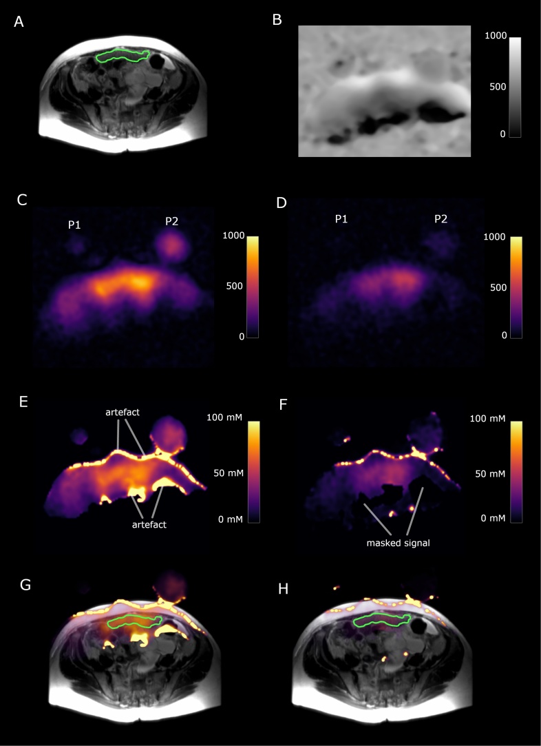
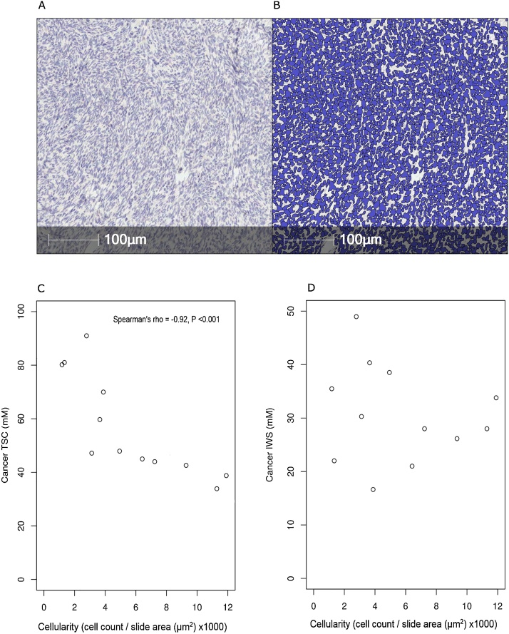
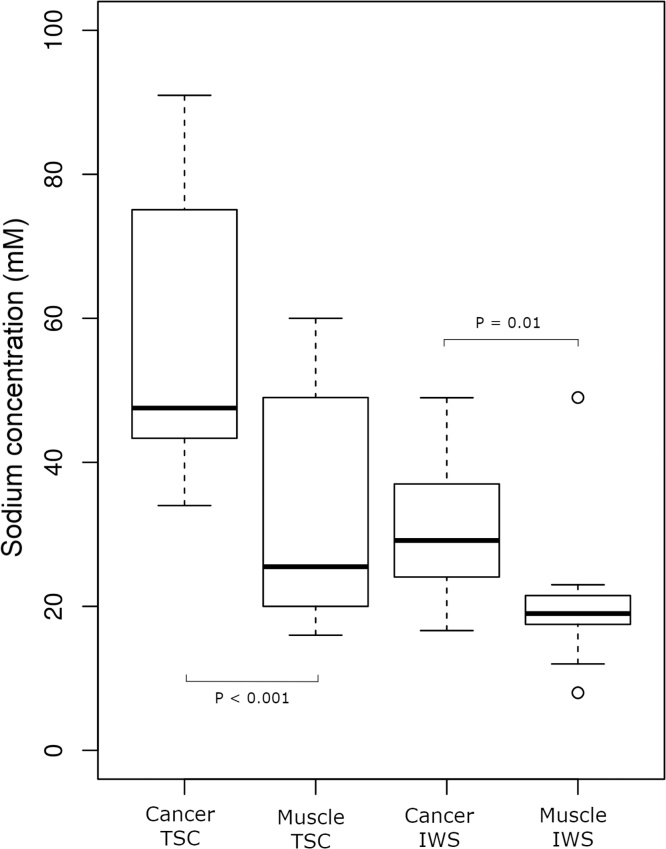
The aim of this study was to assess the feasibility of rapid sodium MRI (Na-MRI) for the imaging of peritoneal cancer deposits in high grade serous ovarian cancer (HGSOC) and to evaluate the relationship of Na-MRI with tumour cellularity. Na-MRI was performed at 3 T on twelve HGSOC patients using a 3D-cones acquisition technique. Tumour biopsies specimens were collected after imaging and cellularity was measured from histology. Total Na-MRI scan time for each patient was approximately 11 min. At an isotropic resolution of 5.6 mm, signal-to-noise ratios (SNRs) of 82.2 ± 15.3 and 15.1 ± 7.1 (mean ± standard deviation) were achieved for imaging of tumour tissue sodium concentration (TSC) and intracellular weighted sodium concentration (IWS) respectively. Tumour TSC and IWS concentrations were: 56.8 ± 19.1 mM and 30.8 ± 9.2 mM respectively and skeletal muscle TSC and IWS concentrations were 33.2 ± 16.3 mM and 20.5 ± 9.9 mM respectively. There were significant sodium concentration differences between cancer and skeletal muscle, Wilcoxon signed-rank test, < 0.001 for TSC and = 0.01 for IWS imaging. Tumour cellularity displayed a strong negative correlation with TSC, Spearman's rho = -0.92, < 0.001, but did not correlate with IWS. This study demonstrates that Na-MRI using 3D-cones can rapidly assess sodium concentration in peritoneal deposits of HGSOC and that TSC may serve as a biomarker of tumour cellularity.
本研究的目的是评估快速钠磁共振成像(Na-MRI)用于高级别浆液性卵巢癌(HGSOC)腹膜癌转移灶成像的可行性,并评估Na-MRI与肿瘤细胞密度的关系。使用3D圆锥采集技术在3T磁场下对12例HGSOC患者进行了Na-MRI检查。成像后采集肿瘤活检标本,并通过组织学测量细胞密度。每位患者的总Na-MRI扫描时间约为11分钟。在各向同性分辨率为5.6mm时,肿瘤组织钠浓度(TSC)成像和细胞内加权钠浓度(IWS)成像的信噪比(SNR)分别为82.2±15.3和15.1±7.1(平均值±标准差)。肿瘤TSC和IWS浓度分别为:56.8±19.1mM和30.8±9.2mM,骨骼肌TSC和IWS浓度分别为33.2±16.3mM和20.5±9.9mM。癌症组织与骨骼肌之间存在显著的钠浓度差异,Wilcoxon符号秩检验显示,TSC成像的P<0.001,IWS成像的P = 0.01。肿瘤细胞密度与TSC呈强负相关,Spearman相关系数rho = -0.92,P<0.001,但与IWS无相关性。本研究表明,使用3D圆锥采集技术的Na-MRI可以快速评估HGSOC腹膜转移灶中的钠浓度,并且TSC可能作为肿瘤细胞密度的生物标志物。