Ishizaka Takuya, Nagano Kazuya, Tasaki Ikkei, Tao Hong, Gao Jian-Qing, Harada Kazuo, Hirata Kazumasa, Saito Shigeru, Tsujino Hirofumi, Higashisaka Kazuma, Tsutsumi Yasuo
Graduate School of Pharmaceutical Sciences, Osaka University, 1-6 Yamadaoka, Suita, Osaka, 565-0871, Japan.
Graduate School of Medicine, Osaka University, 2-2 Yamadaoka, Suita, Osaka, 565-0871, Japan.
Nanoscale Res Lett. 2019 May 28;14(1):180. doi: 10.1186/s11671-019-3016-9.
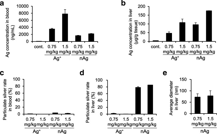
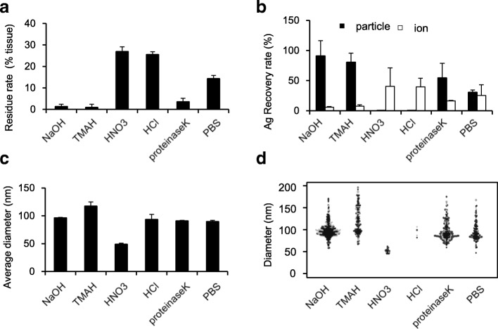
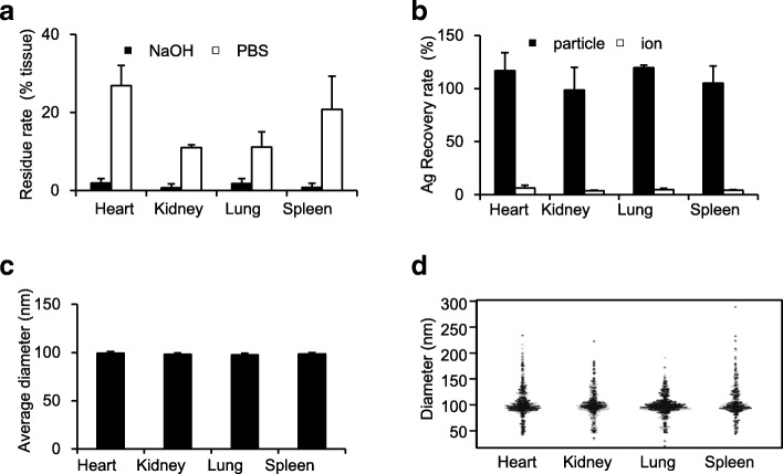
The prevalent use of engineered nanoparticles (ENPs) has increased our exposure to these particles. The current available analytical techniques fail to simultaneously quantify and analyze the physical properties of ENPs in biological tissues. Therefore, new methods are required to evaluate the exposure conditions to ENPs. Single particle inductively coupled plasma-mass spectrometry (sp-ICP-MS) is an attractive approach that can perform quantitative and qualitative analyses of ENPs. However, the application of this approach for biological samples is limited because of the lack of pretreatment methods for effectively recovering ENPs from biological tissues. In this study, we evaluated various pretreatment methods and identified the optimal pretreatment conditions for sp-ICP-MS analyses of ENPs in biological tissues using silver nanoparticles (nAg) as a model. We screened five reagents as pretreatment solvents (sodium hydroxide, tetramethylammonium hydroxide, nitric acid, hydrochloric acid, and proteinase K). Our results showed that treatment with sodium hydroxide was optimal for detecting nAg in the mouse liver. Moreover, this pretreatment method can be applied to other organs, such as the heart, lung, spleen, and kidney. Finally, we evaluated the applicability of this method by analyzing the quantity and physical properties of silver in the mouse blood and liver, after intravenous administration of nAg or silver ion. Our sp-ICP-MS method revealed that nAg administered into the blood was partially ionized and tended to be distributed in the particle form (approximately 80%) in the liver and in ionic form (approximately 95%) in the blood. In conclusion, we optimized pretreatment strategies for sp-ICP-MS evaluation of ENPs in biological tissues and demonstrated its applicability by evaluating the changes in the physical properties of nAg in the liver and blood. We also showed that partial changes from the particle form to the ionic form of nAg influences their kinetics and distribution when administered to mice.
工程纳米颗粒(ENPs)的广泛使用增加了我们对这些颗粒的接触。目前可用的分析技术无法同时量化和分析生物组织中ENPs的物理性质。因此,需要新的方法来评估ENPs的暴露条件。单颗粒电感耦合等离子体质谱法(sp-ICP-MS)是一种有吸引力的方法,可对ENPs进行定量和定性分析。然而,由于缺乏从生物组织中有效回收ENPs的预处理方法,该方法在生物样品中的应用受到限制。在本研究中,我们评估了各种预处理方法,并以银纳米颗粒(nAg)为模型,确定了用于生物组织中ENPs的sp-ICP-MS分析的最佳预处理条件。我们筛选了五种试剂作为预处理溶剂(氢氧化钠、四甲基氢氧化铵、硝酸、盐酸和蛋白酶K)。我们的结果表明,用氢氧化钠处理最适合检测小鼠肝脏中的nAg。此外,这种预处理方法可应用于其他器官,如心脏、肺、脾和肾。最后,我们通过分析静脉注射nAg或银离子后小鼠血液和肝脏中银的含量和物理性质,评估了该方法的适用性。我们的sp-ICP-MS方法显示,注入血液中的nAg部分被离子化,在肝脏中倾向于以颗粒形式分布(约80%),在血液中以离子形式分布(约95%)。总之,我们优化了用于生物组织中ENPs的sp-ICP-MS评估的预处理策略,并通过评估肝脏和血液中nAg物理性质的变化证明了其适用性。我们还表明,nAg从颗粒形式到离子形式的部分变化会影响其注入小鼠后的动力学和分布。