Bugaj Olga, Zieliński Jacek, Kusy Krzysztof, Kantanista Adam, Wieliński Dariusz, Guzik Przemysław
Department of Athletics, Strength and Conditioning, Poznań University of Physical Education, Poznań, Poland.
Department of Sport Kinesiology, Poznań University of Physical Education, Poznań, Poland.
Front Physiol. 2019 May 15;10:600. doi: 10.3389/fphys.2019.00600. eCollection 2019.
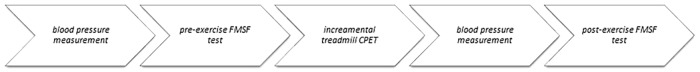
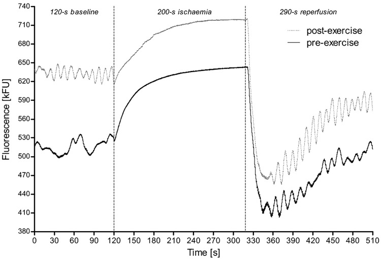
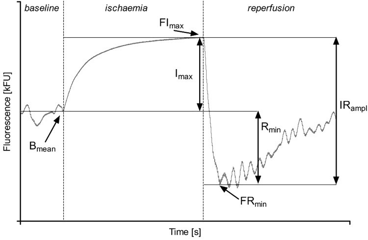
Reduced nicotinamide adenine dinucleotide (NADH) is synthesized in the cellular nucleus, cytoplasm and mitochondria but oxidized into NAD almost exclusively in mitochondria. Activation of human skin by the 340 nm ultraviolet light triggers natural fluorescence at the light length of 460 nm, which intensity is proportional to the skin NADH content. This phenomenon is used by the Flow Mediated Skin Fluorescence (FMSF) which measures changes in the skin NADH content during transient ischemia and reperfusion. We examined the effects of exercise to exhaustion on the skin changes of NADH in response to 200 s forearm ischemia and reperfusion in 121 highly trained athletes (94 men and 27 women, long-distance running, triathlon, taekwondo, rowing, futsal, sprint running, fencing, and tennis). We found that exercise until exhaustion changes the skin content of NADH, modifies NADH turnover at rest, during ischemia and reperfusion in the most superficial living skin cells. Compared to the pre-exercise, there were significant increases in: mean fluorescence recorded during rest as the baseline value ( ) ( < 0.001), the maximal fluorescence that increased above the baseline during controlled forearm ischemia (FI) ( < 0.001, only in men), the minimal fluorescence after decreasing below the baseline during reperfusion (FR) ( < 0.001 men; < 0.01 women) and the difference between and FR ( ) ( < 0.01), and reductions in the difference between FI and ( ) ( < 0.001) and /IR ratio (CI) ( < 0.001) after the incremental exercise test. There was no statistical difference between pre- and post-exercise the maximal range of the fluorescence change during ischemia and reperfusion (IR) In conclusion, exercise to exhaustion modifies the skin NADH content at rest, during ischemia and reperfusion as well as the magnitude of changes in the NADH caused by ischemia and reperfusion. Our findings suggest that metabolic changes in the skin NADH accompanying exercise extend beyond muscles and affect other cells and organs.
还原型烟酰胺腺嘌呤二核苷酸(NADH)在细胞核、细胞质和线粒体中合成,但几乎仅在线粒体中氧化为NAD。340纳米紫外线对人体皮肤的激活会在460纳米波长处引发自然荧光,其强度与皮肤NADH含量成正比。流动介导的皮肤荧光(FMSF)利用了这一现象,该技术可测量短暂性缺血和再灌注期间皮肤NADH含量的变化。我们研究了力竭运动对121名高水平运动员(94名男性和27名女性,包括长跑、铁人三项、跆拳道、赛艇、五人制足球、短跑、击剑和网球运动员)在200秒前臂缺血和再灌注后皮肤NADH变化的影响。我们发现,力竭运动改变了皮肤NADH含量,改变了最表层活皮肤细胞在静息、缺血和再灌注期间的NADH周转率。与运动前相比,静息时记录的平均荧光作为基线值显著增加(P<0.001),在控制性前臂缺血(FI)期间高于基线的最大荧光显著增加(P<0.001,仅在男性中),再灌注(FR)期间低于基线后最小荧光显著增加(男性P<0.001;女性P<0.01)以及FI和FR之间的差值显著增加(P<0.01),递增运动试验后,FI和基线之间的差值(P<0.001)以及I/R比值(CI)(P<0.001)降低。运动前后缺血和再灌注期间荧光变化的最大范围无统计学差异(IR)。总之,力竭运动改变了静息、缺血和再灌注期间皮肤NADH含量以及缺血和再灌注引起的NADH变化幅度。我们的研究结果表明,运动伴随的皮肤NADH代谢变化不仅限于肌肉,还会影响其他细胞和器官。