• 文献检索
  • 文档翻译
  • 深度研究
  • 学术资讯
  • Suppr Zotero 插件Zotero 插件
  • 邀请有礼
  • 套餐&价格
  • 历史记录
应用&插件
Suppr Zotero 插件Zotero 插件浏览器插件Mac 客户端Windows 客户端微信小程序
定价
高级版会员购买积分包购买API积分包
服务
文献检索文档翻译深度研究API 文档MCP 服务
关于我们
关于 Suppr公司介绍联系我们用户协议隐私条款
关注我们

Suppr 超能文献

核心技术专利:CN118964589B侵权必究
粤ICP备2023148730 号-1Suppr @ 2026

文献检索

告别复杂PubMed语法,用中文像聊天一样搜索,搜遍4000万医学文献。AI智能推荐,让科研检索更轻松。

立即免费搜索

文件翻译

保留排版,准确专业,支持PDF/Word/PPT等文件格式,支持 12+语言互译。

免费翻译文档

深度研究

AI帮你快速写综述,25分钟生成高质量综述,智能提取关键信息,辅助科研写作。

立即免费体验

散斑调制可实现高分辨率宽场人脑肿瘤边界检测和活体小鼠神经成像。

Speckle modulation enables high-resolution wide-field human brain tumor margin detection and in vivo murine neuroimaging.

机构信息

Stanford University Department of Structural Biology, Stanford, CA, 94304, USA.

Stanford University Department of Neurosurgery, Stanford, CA, 94304, USA.

出版信息

Sci Rep. 2019 Jul 17;9(1):10388. doi: 10.1038/s41598-019-45902-9.

DOI:10.1038/s41598-019-45902-9
PMID:31316099
原文链接:https://pmc.ncbi.nlm.nih.gov/articles/PMC6637128/
Abstract

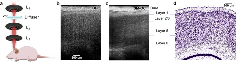
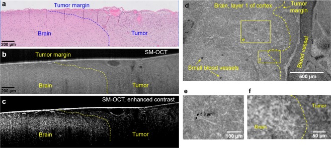
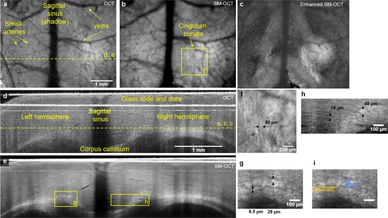
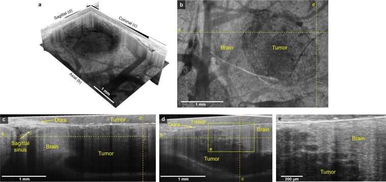
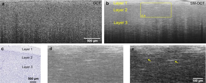
Current in vivo neuroimaging techniques provide limited field of view or spatial resolution and often require exogenous contrast. These limitations prohibit detailed structural imaging across wide fields of view and hinder intraoperative tumor margin detection. Here we present a novel neuroimaging technique, speckle-modulating optical coherence tomography (SM-OCT), which allows us to image the brains of live mice and ex vivo human samples with unprecedented resolution and wide field of view using only endogenous contrast. The increased visibility provided by speckle elimination reveals white matter fascicles and cortical layer architecture in brains of live mice. To our knowledge, the data reported herein represents the highest resolution imaging of murine white matter structure achieved in vivo across a wide field of view of several millimeters. When applied to an orthotopic murine glioblastoma xenograft model, SM-OCT readily identifies brain tumor margins with resolution of approximately 10 μm. SM-OCT of ex vivo human temporal lobe tissue reveals fine structures including cortical layers and myelinated axons. Finally, when applied to an ex vivo sample of a low-grade glioma resection margin, SM-OCT is able to resolve the brain tumor margin. Based on these findings, SM-OCT represents a novel approach for intraoperative tumor margin detection and in vivo neuroimaging.

摘要

目前的活体神经影像学技术提供的视野或空间分辨率有限,并且通常需要外源性对比。这些限制阻碍了在宽视场范围内进行详细的结构成像,并阻碍了术中肿瘤边界的检测。在这里,我们提出了一种新的神经影像学技术,即散斑调制光学相干断层扫描(SM-OCT),它仅使用内源性对比,就可以以前所未有的分辨率和宽视场对活鼠和离体人样本进行成像。散斑消除提供的更高可见度揭示了活鼠大脑中的白质束和皮质层结构。据我们所知,本文报告的数据代表了在活体中在几毫米的宽视场范围内实现的最高分辨率的鼠类白质结构成像。当应用于原位鼠胶质母细胞瘤异种移植模型时,SM-OCT 可以轻松识别分辨率约为 10μm 的脑肿瘤边界。离体人颞叶组织的 SM-OCT 显示出精细的结构,包括皮质层和有髓轴突。最后,当应用于低级别胶质瘤切除边界的离体样本时,SM-OCT 能够分辨出脑肿瘤边界。基于这些发现,SM-OCT 代表了一种用于术中肿瘤边界检测和活体神经影像学的新方法。

https://cdn.ncbi.nlm.nih.gov/pmc/blobs/5de2/6637128/0cfd6f5b8509/41598_2019_45902_Fig5_HTML.jpg
https://cdn.ncbi.nlm.nih.gov/pmc/blobs/5de2/6637128/6930d78efb08/41598_2019_45902_Fig1_HTML.jpg
https://cdn.ncbi.nlm.nih.gov/pmc/blobs/5de2/6637128/a7baf770c38e/41598_2019_45902_Fig2_HTML.jpg
https://cdn.ncbi.nlm.nih.gov/pmc/blobs/5de2/6637128/c10e1bb1949f/41598_2019_45902_Fig3_HTML.jpg
https://cdn.ncbi.nlm.nih.gov/pmc/blobs/5de2/6637128/1cef43d20db2/41598_2019_45902_Fig4_HTML.jpg
https://cdn.ncbi.nlm.nih.gov/pmc/blobs/5de2/6637128/0cfd6f5b8509/41598_2019_45902_Fig5_HTML.jpg

相似文献

1
Speckle modulation enables high-resolution wide-field human brain tumor margin detection and in vivo murine neuroimaging.散斑调制可实现高分辨率宽场人脑肿瘤边界检测和活体小鼠神经成像。
Sci Rep. 2019 Jul 17;9(1):10388. doi: 10.1038/s41598-019-45902-9.
2
Spatiotemporal Tracking of Brain-Tumor-Associated Myeloid Cells through Optical Coherence Tomography with Plasmonic Labeling and Speckle Modulation.通过等离子体标记和散斑调制的光相干断层扫描对脑肿瘤相关髓样细胞进行时空追踪。
ACS Nano. 2019 Jul 23;13(7):7985-7995. doi: 10.1021/acsnano.9b02656. Epub 2019 Jul 1.
3
Laser brain cancer surgery in a xenograft model guided by optical coherence tomography.光学相干断层扫描引导下的异种移植模型中的激光脑癌手术。
Theranostics. 2019 May 26;9(12):3555-3564. doi: 10.7150/thno.31811. eCollection 2019.
4
Application of multi-wavelength technique for photoacoustic imaging to delineate tumor margins during maximum-safe resection of glioma: A preliminary simulation study.多波长技术在最大安全切除脑胶质瘤过程中的光声成像中的应用:初步模拟研究。
J Clin Neurosci. 2019 Dec;70:242-246. doi: 10.1016/j.jocn.2019.08.040. Epub 2019 Aug 30.
5
Quantification of attenuation and speckle features from endoscopic OCT images for the diagnosis of human brain glioma.从内镜 OCT 图像中量化衰减和散斑特征以诊断人脑胶质瘤。
Sci Rep. 2024 May 10;14(1):10722. doi: 10.1038/s41598-024-61292-z.
6
AI-Assisted Detection of Human Glioma Infiltration Using a Novel Computational Method for Optical Coherence Tomography.利用一种新型的光学相干断层扫描计算方法辅助检测人脑胶质瘤浸润。
Clin Cancer Res. 2019 Nov 1;25(21):6329-6338. doi: 10.1158/1078-0432.CCR-19-0854. Epub 2019 Jul 17.
7
Feasibility evaluation of micro-optical coherence tomography (μOCT) for rapid brain tumor type and grade discriminations: μOCT images versus pathology.微光学相干断层扫描(μOCT)在快速脑肿瘤类型和分级鉴别中的可行性评估:μOCT 图像与病理学比较。
BMC Med Imaging. 2019 Dec 30;19(1):102. doi: 10.1186/s12880-019-0405-6.
8
Speckle-modulating optical coherence tomography in living mice and humans.活体小鼠和人类中的斑点调制光学相干断层扫描。
Nat Commun. 2017 Jun 20;8:15845. doi: 10.1038/ncomms15845.
9
Theranostic OCT microneedle for fast ultrahigh-resolution deep-brain imaging and efficient laser ablation in vivo.用于快速超高分辨率深脑成像和体内高效激光烧蚀的治疗性 OCT 微针。
Sci Adv. 2020 Apr 10;6(15):eaaz9664. doi: 10.1126/sciadv.aaz9664. eCollection 2020 Apr.
10
Delineation of three-dimensional tumor margins based on normalized absolute difference mapping via volumetric optical coherence tomography.基于容积光学相干断层扫描的归一化绝对差映射勾画三维肿瘤边界。
Sci Rep. 2024 Apr 5;14(1):7984. doi: 10.1038/s41598-024-56239-3.

引用本文的文献

1
Research progress on the use of the optical coherence tomography system for the diagnosis and treatment of central nervous system tumors.光学相干断层扫描系统在中枢神经系统肿瘤诊断与治疗中的应用研究进展
Ibrain. 2024 Nov 22;11(1):3-18. doi: 10.1002/ibra.12184. eCollection 2025 Spring.
2
Optical coherence tomography (OCT) and OCT angiography: Technological development and applications in brain science.光学相干断层扫描(OCT)与OCT血管造影:技术发展及其在脑科学中的应用
Theranostics. 2025 Jan 1;15(1):122-140. doi: 10.7150/thno.97192. eCollection 2025.
3
Noninvasive virtual biopsy using micro-registered optical coherence tomography (OCT) in human subjects.

本文引用的文献

1
as-PSOCT: Volumetric microscopic imaging of human brain architecture and connectivity.基于复合散射光学相干断层成像术(as-PSOCT)的人脑结构与连接的容积微观成像
Neuroimage. 2018 Jan 15;165:56-68. doi: 10.1016/j.neuroimage.2017.10.012. Epub 2017 Oct 7.
2
Rethinking childhood ependymoma: a retrospective, multi-center analysis reveals poor long-term overall survival.重新思考儿童室管膜瘤:回顾性多中心分析显示长期总体生存率较差。
J Neurooncol. 2017 Oct;135(1):201-211. doi: 10.1007/s11060-017-2568-8. Epub 2017 Jul 21.
3
Speckle-modulating optical coherence tomography in living mice and humans.
利用微配准光学相干断层扫描(OCT)对人体进行无创虚拟活检。
Sci Adv. 2024 Apr 12;10(15):eadi5794. doi: 10.1126/sciadv.adi5794. Epub 2024 Apr 10.
4
Research progress on the application of optical coherence tomography in the field of oncology.光学相干断层扫描在肿瘤学领域应用的研究进展
Front Oncol. 2022 Jul 25;12:953934. doi: 10.3389/fonc.2022.953934. eCollection 2022.
5
Intravital 3D visualization and segmentation of murine neural networks at micron resolution.微米分辨率下的小鼠神经网络的活体 3D 可视化和分割。
Sci Rep. 2022 Jul 30;12(1):13130. doi: 10.1038/s41598-022-14450-0.
6
Applying machine learning to optical coherence tomography images for automated tissue classification in brain metastases.将机器学习应用于光学相干断层扫描图像,实现脑转移瘤的自动组织分类。
Int J Comput Assist Radiol Surg. 2021 Sep;16(9):1517-1526. doi: 10.1007/s11548-021-02412-2. Epub 2021 May 30.
7
Capability of physically reasonable OCT-based differentiation between intact brain tissues, human brain gliomas of different WHO grades, and glioma model 101.8 from rats.基于光学相干断层扫描(OCT)在完整脑组织、不同世界卫生组织(WHO)分级的人脑胶质瘤以及大鼠胶质瘤模型101.8之间进行物理合理区分的能力。
Biomed Opt Express. 2020 Oct 28;11(11):6780-6798. doi: 10.1364/BOE.409692. eCollection 2020 Nov 1.
8
Longitudinal in-vivo OCM imaging of glioblastoma development in the mouse brain.小鼠脑内胶质母细胞瘤发展的纵向活体光学相干断层扫描成像
Biomed Opt Express. 2020 Aug 12;11(9):5003-5016. doi: 10.1364/BOE.400723. eCollection 2020 Sep 1.
9
Angular compounding for speckle reduction in optical coherence tomography using geometric image registration algorithm and digital focusing.基于几何图像配准算法和数字聚焦的光学相干断层扫描散斑减少的角度复合。
Sci Rep. 2020 Feb 5;10(1):1893. doi: 10.1038/s41598-020-58454-0.
10
Aperture phase modulation with adaptive optics: a novel approach for speckle reduction and structure extraction in optical coherence tomography.基于自适应光学的孔径相位调制:光学相干断层扫描中减少散斑和提取结构的新方法。
Biomed Opt Express. 2019 Jan 15;10(2):552-570. doi: 10.1364/BOE.10.000552. eCollection 2019 Feb 1.
活体小鼠和人类中的斑点调制光学相干断层扫描。
Nat Commun. 2017 Jun 20;8:15845. doi: 10.1038/ncomms15845.
4
Robust and fast characterization of OCT-based optical attenuation using a novel frequency-domain algorithm for brain cancer detection.使用基于 OCT 的新型频域算法对脑癌进行快速、稳健的光学衰减特征分析。
Sci Rep. 2017 Mar 22;7:44909. doi: 10.1038/srep44909.
5
Multimodal optical analysis discriminates freshly extracted human sample of gliomas, metastases and meningiomas from their appropriate controls.多模态光学分析可区分新鲜提取的脑胶质瘤、转移瘤和脑膜瘤患者样本与其相应对照样本。
Sci Rep. 2017 Feb 2;7:41724. doi: 10.1038/srep41724.
6
Extended-focus optical coherence microscopy for high-resolution imaging of the murine brain.用于小鼠大脑高分辨率成像的扩展焦深光学相干显微镜
Biomed Opt Express. 2016 Oct 5;7(11):4400-4414. doi: 10.1364/BOE.7.004400. eCollection 2016 Nov 1.
7
Live volumetric (4D) visualization and guidance of in vivo human ophthalmic surgery with intraoperative optical coherence tomography.术中光学相干断层扫描引导活体人眼手术的实时容积(4D)可视化。
Sci Rep. 2016 Aug 19;6:31689. doi: 10.1038/srep31689.
8
Comprehensive cellular-resolution atlas of the adult human brain.成年人类大脑的全细胞分辨率图谱。
J Comp Neurol. 2016 Nov 1;524(16):3127-481. doi: 10.1002/cne.24080.
9
Third harmonic generation imaging for fast, label-free pathology of human brain tumors.用于人脑肿瘤快速、无标记病理学分析的三次谐波产生成像
Biomed Opt Express. 2016 Apr 18;7(5):1889-904. doi: 10.1364/BOE.7.001889. eCollection 2016 May 1.
10
Polarization sensitive optical coherence microscopy for brain imaging.用于脑成像的偏振敏感光学相干显微镜
Opt Lett. 2016 May 15;41(10):2213-6. doi: 10.1364/OL.41.002213.