Bermúdez Vicente, Tenconi Paula Estefanía, Giusto Norma María, Mateos Melina Valeria
Instituto de Investigaciones Bioquímicas de Bahía Blanca (INIBIBB), Consejo Nacional de Investigaciones Científicas y Técnicas (CONICET), Bahía Blanca, Argentina.
Departamento de Biología, Bioquímica y Farmacia (DBByF), Universidad Nacional del Sur (UNS), Bahía Blanca, Argentina.
Front Cell Neurosci. 2019 Apr 24;13:154. doi: 10.3389/fncel.2019.00154. eCollection 2019.
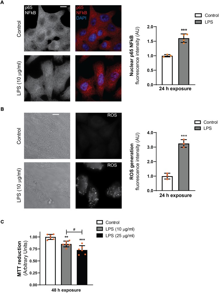
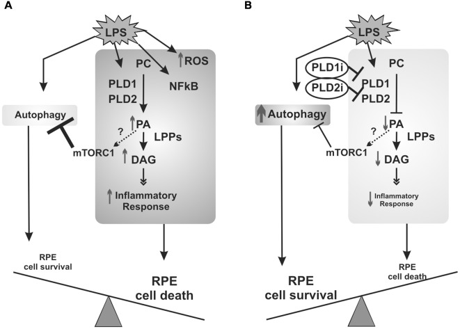
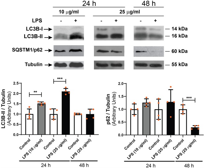
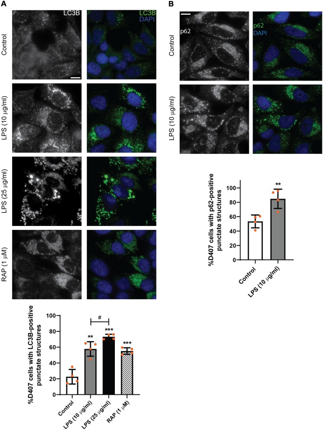
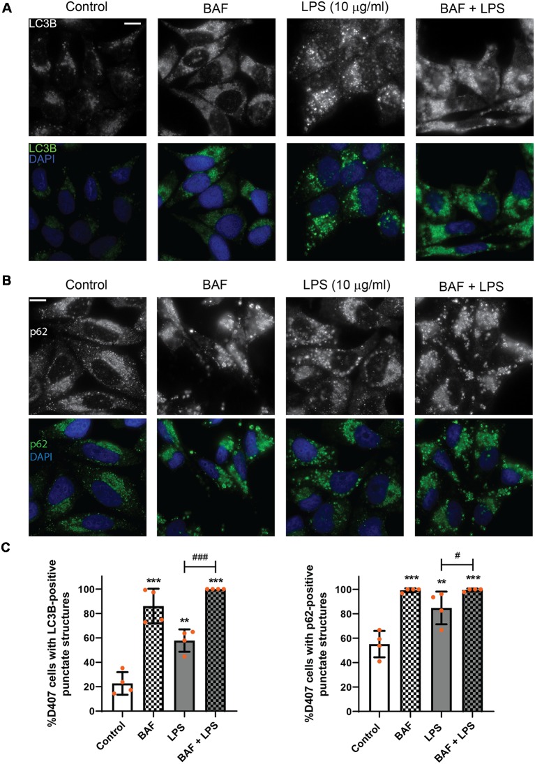
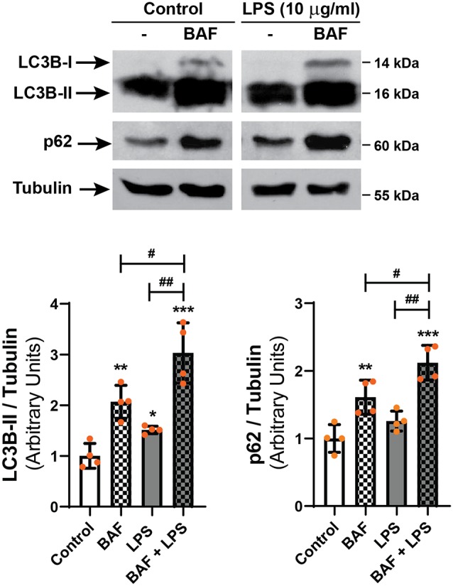
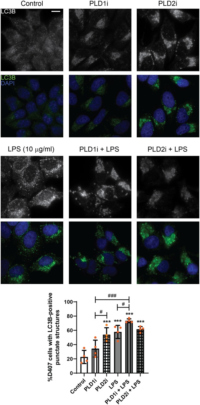
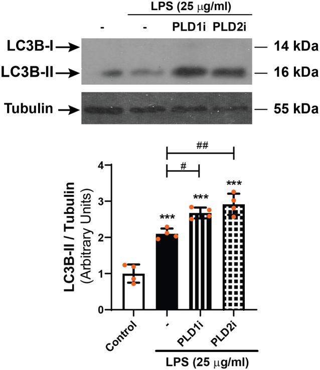
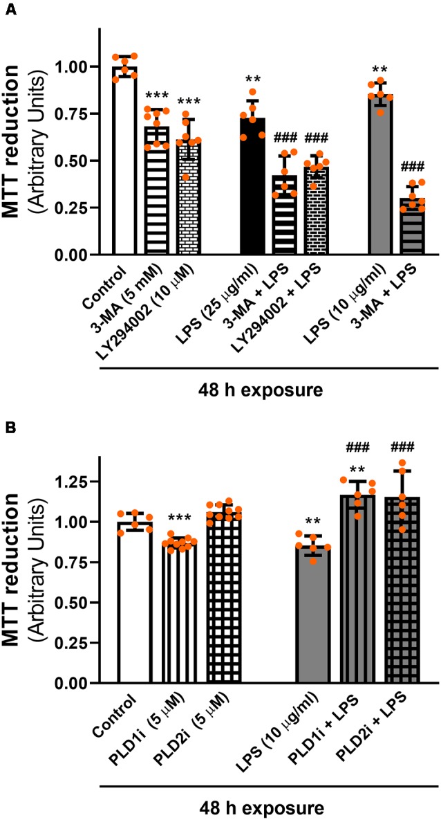
Inflammation and oxidative stress are common factors involved in the pathogenesis of retinal diseases, such as aged-related macular degeneration (AMD) and diabetic retinopathy (DR). Autophagy is a catabolic process essential to cell survival in response to stress. This process is highly active in retinal pigment epithelium (RPE) cells. Our previous findings demonstrated that lipopolysaccharide (LPS) induces an inflammatory response of RPE cells that implies classical phospholipases D (PLD1 and 2) activation, cyclooxygenase-2 (COX-2) expression, prostaglandin E (PGE) production and reduced cell viability. In this work, we studied the autophagic process and its modulation by the PLD pathway in D407 and ARPE-19 RPE cells exposed to LPS. LPS (10 μg/ml or 25 μg/ml) exposure for 24 h increased light chain 3B-II (LC3B-II) content (an autophagy marker) and LC3B-positive punctate structures in both RPE cell lines studied. Next, the drug bafilomycin A (BAF, 50 nM) was used to block the autophagic flux. In cells pre-incubated with BAF, LC3B-II and sequestosome 1 (SQSTM1/p62) levels and autophagosome-like structures were increased by LPS, demonstrating that the inflammatory injury increases the autophagic process in RPE cells. To study the role of the PLD pathway, cells were pre-incubated for 1 h with selective PLD1 (VU0359595) or PLD2 (VU0285655-1) inhibitors prior to LPS addition. Under control condition, LC3B-positive punctate structures were increased in cells pre-incubated with PLD2 inhibitor while with PLD1 inhibitor were increased in cells exposed to LPS. MTT reduction assays showed that early autophagy inhibitors, 3-methyladenin (3-MA) or LY294002, enhanced the loss in cell viability induced by LPS exposure for 48 h. On the contrary, the inhibition of PLD1 and PLD2 prevented the loss in cell viability induced by LPS. In conclusion, our results show that even though LPS treatment promotes an inflammatory response in RPE cells, it also triggers the activation of the autophagic process which in turn may serve as a protective mechanism for the cells. In addition, we demonstrate that the PLD pathway modulates the autophagic process in RPE cells. Our findings contribute to the knowledge of the molecular basis of retinal inflammatory and degenerative diseases and open new avenues for potential therapeutic exploration.
炎症和氧化应激是视网膜疾病发病机制中常见的因素,如年龄相关性黄斑变性(AMD)和糖尿病视网膜病变(DR)。自噬是细胞在应激状态下生存所必需的分解代谢过程。这一过程在视网膜色素上皮(RPE)细胞中高度活跃。我们之前的研究结果表明,脂多糖(LPS)可诱导RPE细胞的炎症反应,这意味着经典磷脂酶D(PLD1和2)被激活、环氧合酶-2(COX-2)表达、前列腺素E(PGE)产生以及细胞活力降低。在这项研究中,我们研究了暴露于LPS的D407和ARPE-19 RPE细胞中的自噬过程及其受PLD途径的调节。LPS(10μg/ml或25μg/ml)处理24小时可增加所研究的两种RPE细胞系中的轻链3B-II(LC3B-II)含量(一种自噬标志物)和LC3B阳性点状结构。接下来,使用巴弗洛霉素A(BAF,50 nM)药物来阻断自噬流。在预先用BAF孵育的细胞中,LPS可增加LC3B-II和隔离小体1(SQSTM1/p62)水平以及自噬体样结构,这表明炎症损伤会增加RPE细胞中的自噬过程。为了研究PLD途径的作用,在添加LPS之前,细胞先用选择性PLD1(VU0359595)或PLD2(VU0285655-1)抑制剂孵育1小时。在对照条件下,预先用PLD2抑制剂孵育的细胞中LC3B阳性点状结构增加,而预先用PLD1抑制剂孵育的细胞在暴露于LPS时LC3B阳性点状结构增加。MTT还原试验表明,早期自噬抑制剂3-甲基腺嘌呤(3-MA)或LY294002可增强LPS暴露48小时诱导的细胞活力丧失。相反,抑制PLD1和PLD2可防止LPS诱导的细胞活力丧失。总之,我们的结果表明,尽管LPS处理会促进RPE细胞中的炎症反应,但它也会触发自噬过程的激活,这反过来可能作为细胞的一种保护机制。此外,我们证明PLD途径可调节RPE细胞中的自噬过程。我们的研究结果有助于了解视网膜炎症和退行性疾病的分子基础,并为潜在的治疗探索开辟新途径。