Han Lv, Na Zeng, Chunli Liu, Yuchen Chen, Pengfei Zhao, Hao Wang, Xu Cheng, Peng Zhang, Zheng Wang, Zhenghan Yang, Shusheng Gong, Zhenchang Wang
Department of Radiology, Beijing Friendship Hospital, Capital Medical University, Beijing, China.
National Clinical Research Center for Digestive Diseases, Beijing Friendship Hospital, Capital Medical University, Beijing, China.
Front Neurosci. 2019 Jul 4;13:614. doi: 10.3389/fnins.2019.00614. eCollection 2019.
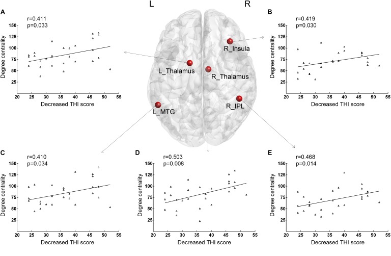
Previous resting-state functional magnetic resonance imaging (fMRI) studies have shown neural connectivity alterations after the treatment of tinnitus. We aim to study the value of the baseline functional connectivity features of neural network nodes to predict outcomes of sound therapy through adjusted narrow band noise. The fMRI data of 27 untreated tinnitus patients and 27 matched healthy controls were analyzed. We calculated the graph-theoretical metric degree centrality (DC) to characterize the functional connectivity of the neural network nodes. Therapeutic outcomes are determined by the changes in the Tinnitus Handicap Inventory (THI) score after a 12-week intervention. The connectivity of 10 brain nodes in tinnitus patients was significantly increased at baseline. The functional connectivity of right insula, inferior parietal lobule (IPL), bilateral thalami, and left middle temporal gyrus was significantly modified with the sound therapy, and such changes correlated with THI changes in tinnitus patients. Receiver operating characteristic curve analyses revealed that the measurements from the five brain regions were effective at classifying improvement after therapy. After age, gender, and education correction, the adjusted area under the curve (AUC) values for the bilateral thalami were the highest (left, 0.745; right, 0.708). Our study further supported the involvement of the fronto-parietal-cingulate network in tinnitus and found that the connectivity of the thalamus at baseline is an object neuroimaging-based indicator to predict clinical outcome of sound therapy through adjusted narrow band noise.
先前的静息态功能磁共振成像(fMRI)研究表明,耳鸣治疗后神经连接性会发生改变。我们旨在研究神经网络节点的基线功能连接特征对于预测通过调整窄带噪声进行声音治疗效果的价值。分析了27名未经治疗的耳鸣患者和27名匹配的健康对照者的fMRI数据。我们计算了图论指标度中心性(DC)来表征神经网络节点的功能连接性。治疗效果通过12周干预后耳鸣障碍量表(THI)评分的变化来确定。耳鸣患者中10个脑节点的连接性在基线时显著增加。右侧岛叶、顶下小叶(IPL)、双侧丘脑和左侧颞中回的功能连接性在声音治疗后有显著改变,且这些变化与耳鸣患者的THI变化相关。受试者工作特征曲线分析显示,来自五个脑区的测量结果在分类治疗后改善情况方面是有效的。在对年龄、性别和教育程度进行校正后,双侧丘脑的校正曲线下面积(AUC)值最高(左侧,0.745;右侧,0.708)。我们的研究进一步支持了额顶扣带网络参与耳鸣的过程,并发现基线时丘脑的连接性是一种基于神经影像学的指标,可用于预测通过调整窄带噪声进行声音治疗的临床效果。